Author Archives: rajani

Immune Cells in COVID-19

Introduction

COVID-19 pandemic spread in almost all, all over the globe countries .Millions have been affected and millions died. The situation necessitate international efforts of medical internists epidemiologists, immunologist, and vaccine technologist .Four main preventive vaccine versions were becoming in hand for human mass vaccination within one year after the pandemic spread. So far concerning immunology of the disease. Scientists early in the pandemic starts to assess humoral immunity suitable for sero-diagnosis and sero-therapy of the disease as well as tempting and still tempting to developing the COVID-19 valid preventive vaccines. Currently, cell and lymphocyte immunologists take the forefront position in COVID-19 research. They articulate cellular immunology, flow cytometery, single cell RNA sequencing, mass transcriptomics, proteomics and cell-cell inter- actomics for tracing lymphocyte immunotypes and the allied cell type interactions .As well as their roles in; the pathogenesis, immune- pathogenesis, and disease outcome prediction.

Aiming

The objective of the present special issue “IMMUNE CELLS  IN COVID-19” published in IJPC was focusing onto the; ontogeny, biology, molecular biology, immuno-typing of immune cells and their roles in covuid-19.In which the latest information were collected, analyzed and formatted as series of opinion papers.

Scope

Lymphoid, lympho-myeloid and myeloid cell lineages for lymphocytes, monocytes and granulocytes as well as their mature forms are forming the backbone of human and mammalian immune system. The function of the immune system is mainly to recognize and destroy foreign invaders via a tripartite responses as; innate, immune cross-roads and adaptive immune responses. Immune cells the subject of the present special issue were assigned to these three immune system arms.

List of Contents

Section One: Innate Immunity

  1. Immunology of mononuclear phagocytes system in pulmonary patho-type of COVID-19.*
  2. Section Two: Immune Cross-roads

    1. The interplay of NKT cells in severe sars-cov-2 human infections*
    2. TH17 cells and the intercellular functions in severe, critical, deceased and vaccine of COVID-19*
    3. Section Three: Adaptive Immunity

      1. B Cell immunology of COVID-19*.
      2. Regulatory Lymphocytes in COVID-19*.
      3. 6-MAIT functions in homeostasis,covid-19 infected an covid-19
      4. *The authorship;

        IBRAHIM MS SHNAWA, College of Biotechnology, University of Qasim and Hilla University, College Babylon, Iraq.

Opinion: Immunology of Mononuclear Phagocyte System in Pulmonary Pathotype of COVID-19

Abstract

Mononuclear Phagocyte System MPS has an essential role in all stages of human SARS-COV-2 pulmonary infections. The objective of the present opinion paper was to through a light on the forefront achievements on the immunology of MPS in COVID-19. Single cell mRNA sequencing, single nucleus RNA sequencing, PCR, transcriptomics, flow cytometery, histo and gross pathology were the main assays tempted for assessments through an in-vitro, ex-vivo and in-vivo experimental settings. Monocyte, macrophage, alveolar macrophage, dendritic cells via an increment or decrement shift in number or function could probe the disease severity. MPS cells are either primer infected and lead to serial cellular events ended with severity. Or the epithelial cells found in the micro-environmental continuum with the APS were infected leading to MPS cell infection followed by cell-cell cross-talks, positive loop feedback mechanisms with T lymphocyte and/or MPS cells interferon axis functions. The overall immune response patterns of the lung in severe COVID-19 were; hyper-inflammation, immune impairment, hypoxia and severity terminated with death if not managed at earliest. An immune six point severity index was proposed as a diagnostic battery to be of use in an advance immunology laboratory was suggested. Molecular immune concept of circuit was briefed. MPS immune functions in pulmonary COVID-19 hold the position of double sward beneficial in some functional aspects and deleterious in others.

Keywords

Alveolar, Asymptomatic, Cytometery, Dendritic cell, Flow, Infection, Macrophages, Monocyte

Introduction

Mononuclear phagocyte system MPS take part in the functions of the human immune system both in health and disease. As a system is composed of circulating monocyte and tissue resident forms, the tissue resident forms got different names in different tissue microenvironment as; Glial in brain, alveolar in lungs, Kupffer in liver, osteoclast in bone, dendritic in spleen and other lymph glands and blood stream and Langerhans in skin. Some of which undergoes phase transition as that of glial cells in central nervous system. MPS performed immune functions both in the natural (innate) and adaptive immune responses. In other word they take part in immune cross-roads functions. In general MPS interplay immune functions in viral diseases and have special immune potentials in COVID-19. In health, MPS performed; phagocytosis, antigen presentation, shaping the adaptive immune responses, production of cytokines  and chemokines. While in disease state MPS played a role in the infectious inflammatory processes and in immune tissue  injuries due to an excessive cytokine production insitue in the affected tissue microenvironment [1-3]. In the present opinion tempts were made  to review the immunology of mononuclear phagocyte cell system in pulmonary COVID-19.

Cellular Immunology of MPS

MPS cells originated from the pluripotent stem cells in bone marrow in human adults. From the stem cells, lympho-myeloid progenitor cell line was developed which then differentiated to pro- monoblast, mono-blast, pro-monocyte then to monocyte in blood stream. From blood stream migrate to tissue compartments. During such migration they undergo morphologic and functional changes that fits to the target tissue compartment or organ while migration within blood vessels would not accompanied by morphologic, functional and/or mitogenic changes.

MPS cells are of large sizes and have multiple secondary lysozymes. They are characterized  by  active  endoplasmic reticulum and active Golgi apparatus which means  have  both  active biosynthetic pathways and active secretions with an evident acclimatization to their microenvironment. In lungs MPS express oxidative metabolism, their half-life were ranged between 60 to90 days in various organ/systems. During the inflammatory stimuli, therein  there  are  increments  in  the  in  their  development   in bone marrow and disseminated to various tissues through blood stream. On surface of MPCS cells there are numbers of markers   like:  MHCI,  MHCII,  C1,  C2,  FC,  CD11,  CD18,  CD13,   CD16,

CD17, CD31, and IA. The biophysical characteristics of MPS are; stimulated by bacterial lipo-polysachaaride LPS and mitogenic lectins, adhere to glass, recognize the antigens by TLR receptor. They secrete; hydrolytic enzymes, enzyme  inhibitors,  cytokines,  fat derived factors, complement components, and mirobicidal materials. When activated lymphocyte produce both of; macrophage inhibitory factor MIF it affects inhibition in spread of macrophages and   macrophage   stimulating   factors   which   stimulate   them for pinocytosis, phagocytosis, and induce the appearance of immune associated antigen Ia and assist in antigen presentation. MPS own specialized mechanisms for recognition of different inflammatory stimuli. If the stimulus is microbe, they will evolve number of killing mechanisms. They evolve three highly efficient recognition and clearing mechanisms of immune complexes through phagocytosis. The presence of MPS in continuum of  inflammatory  responses  they indicated either sub-acute or chronic inflammatory state. MPS cells act as; second line defender of human and mammalian body, antigen processing, antigen presentation, cytokine production and recognition of inflammatory responses [1-3].

Cell Molecular Immunology

There are varieties of natural immune protein that have the ability to recognize and detect human infection. These proteins are either soluble or structural entities like; soluble lysosome, complement components or complement receptors. The recognition process is through pattern recognition on the surface of the microbes. Among these receptors are the Toll-like receptor, the TLRs which are proteins of collectin nature that have the ability to recognize certain molecular patterns on the microbial surface. Such recognition molecules are forming strong features of natural (innate) immunity. TLRs are considered as a part of normal immune physiology. In the structural sense TLRs are forming a family of trans-membrane proteins that belongs to a class of animal lectins known as collectin proteins. TLR family is composed of more than ten different receptors. Most of the human and mammalian body tissues express at least one type of TLR. Though all TLRs are expressed onto; macrophages, dendritic cells, mast cells and B cells. TLRs interplayed an array of immune functions like; microbial sensors, cell signaling activators, enhancers for the expression of both; inflammatory and immune response genes, cross- linking of pattern-recognition molecules on the surface of microbe with TLRs act as danger signal to increase the microbicidal activity of phagocytic macrophages and allow them to activate T cells. TLRs made an important link between innate and adaptive immune responses. On TLRs activation, macrophage co-stimulatory molecules will converts macrophage phagocytes into antigen presenting macrophages which able to activate T cells [4-7].

Immunology of MPS in Human Virus Infections

The invading mammalian and human viruses when gain foot- hold in their respective hosts. They are recognized via their surface pattern recognition molecules PRM by the surface TLRs of MPS in blood stream and tissue resident. PRM cross-linked TLRs then virus pinocytosed, or through macro-pinocytosis and/or receptor mediated endocytosis in to the cell interior, Nikitina et al. [2018] the cross-link lead to transform of pinocytosed cell into antigen presenting cells. The processed virus peptides conjugated with MHC molecule and migrate out on the surface of MPS. The antigen presenting MPS will either activate naïve T cell to be Th2 triggering naïve b lymphocytes to grow, proliferate and expand as an effector antibody producing and memory B cells. Or activate naïve T cells to Th1 cells triggering T cells to be effector CD8+ cytotoxic T cells, CD4 T cells and memory T cells. The burden of virus load can be eradicated by the action of cytotoxic T cells or neutralized by the antiviral antibody and/or cleared by the direct action of interferons. The presence of molecular mimicking viral epitope with host tissue cells may initiate through the action of autoantibodies or auto-reactive cells immune tissue injuries terminated by autoimmune diseases. Excessive cytokine production by T cells or MPS also lead to inflammatory and immune responses with consequences of immune tissue injuries. The possible occurrence of viral immunosuppressive epitopes will trigger a state of infectious immunosuppressive conditions [6-9].

Immunology of MPS in SARS-COV-2 Infections

The circulating and tissue resident MPS cells, the monocyte and tissue macrophages participate in all stages of SARS-COV-2 human infection. They contribute to: (i) innate immune reactions (ii) shaping adaptive immune reactions, (iii) comorbidity predisposing to clinical infections, (iv) virus resistance, (v) virus dissemination, (vi) the host factors that determine disease severity, (vii) induction of immune tissue injury, (viii) recovery and (ix) sequalae (Table 1) [10-13].

Table 1: Immune cell deviations in human COVID-19 lungs

Features

Immune Events

References


Molecular

(i) Macrophage infection via antibody dependent receptor mediated endocytosis or pinocytosis

(ii) Amplification of cytokine synthesis and secretion

(iii) pyroptosis

(i) [14]

(ii) [15]

(iii) [14]

Surface markers (i) DCs lack of surface markers

(ii) appearance of an inhibitor surface markers DCs

[16]


Whole cell Immune Deviation

(i) appearance of intermediate phenotypes

(ii) appearance of suppressor phenotypes

(iii) DC-interferon axis

(iv) impaired phase transition in alveolar epithelial cells

(v) Bilateral alveolar macrophage positive feedback loop with T cells

(vi) immune mediated pulmonary fibrosis

(i) [17]

(ii) [17]

(iii) [18]

(iv) [19] (v) [20]

(vi) [21][19]


Molecular inflammatory Events

(i) Infammosome formation

(ii) Hyper-inflammatory responses

(iii) Hypercytokinemia

(iv) Pyroptosis

[22]

[23]

[15]

[11]

[14]

Gross Inflammatory Response outcomes Plogs in all respiratory tracts, transudates and edema [21]

Immuno-Inflammatory Responses

Severe SARS-COV-2 infection induce haemo-phagocytic syndrome due to the infiltration of pro-inflammatory monocytes, a rare condition expressed as an over excerbant inflammatory response due to development of hyper-cytokinemia together with depletion of the adaptive immune compartment which may explain the appearance of sepsis in many severe COVID-19 cases Gomez-Rail [23]. Macrophage activation syndrome MAS is a condition of systemic hyper- inflammation and often be noted in infection and malfunctioning. It is typified by marked up-regulated expression of pro-inflammatory cytokines. This sort of strong inflammation results in severe tissue injury. Macrophage within MAS state produce high amount of pro- inflammatory cytokine upon stimulation.  Inflammation  is  known to destruct the balance between coagulation and fibrionolysis [14]. The inflammatory cytokines TNFalpha and IL1 instruct macrophage and monocytes to produce tissue factor TF. TF activate coagulation while IL1 anIL6 increase the production of plasmalogin activation inhibitor. Hence, overproduction of inflammatory cytokines along with MAS also promotes intravascular coagulation Otsuka and Senio [15]. Dys-regulated inflammatory syndrome DIS. DIS is generated by mononuclear phagocytes (a rich source of pro-inflammatory cytokines) upon encounter of the virus within the tissue continuum via two stage activation mechanisms which is not specific to the initiating virus. This is relevant to the case of SARS-COV-2 virus infection were age and predisposing comorbidities enhances the risk of severe outcome due to DIS [11].

Human severe SARS-COV-2 pulmonary infection leads to inflammation and tissue destruction with a consequence of an immune mediated fibrosis which remains even in to convalescent phase. In a group of severe pulmonary infected patients with COVID-19. IA aided CT scan were used to score fibrosis via fibrosis index IF. Twelve patients with severe COVID-19 were investigated for IF, they were sub-grouped into IFlo and IF hi. Mononuclear cell were collected from those patients and investigated by single cell RNA sequencing, IF hi group have shown low mononuclear phagocytic cell, low IFN gene profiling as compared to that of IFlo subgroup   of patients. Mononuclear phagocyte could probe the prognosis of immune mediated ling fibrosis [16-21].

Monocyte Responses

In an in-vivo study setting SARS-COV-2 infection sensed by monocyte and macrophages, such sensation forms the inflammosomes that activate caspase I and gasdermin D leading to inflammatory cell death, the pyroptosis and release of potent inflammatory mediators. About 6% of blood monocyte of COVID-19 patients are infected with SARS-COV-2 virus. This virus infection of monocyte depends on the uptake of antibody-opsoinzed virus by FC gamma receptors. The internalized virus begins to replicate within the infected monocyte but infection is aborted and the infectious virus was not detected in the culture supernatant of the infected monocyte. Instead the infected monocyte undergoes pyroptosis mediated by activation of NLRP3 and AIM2 inflammosomes, caspase I and gasdermin D. In same culture settings, the addition of the COVID-19 vaccinee plasma does not promote AB dependent monocyte infection. Moreover tissue resident macrophages but not the infected epithelium and endothelium from lung autopsies from the deceased patients with COVID-19 have activated inflammosomes. The overall of findings suggest that ab-dependent SARS-COV-2 uptake by monocytes and macrophages triggers inflammatory cell death that abort the products of the infectious virus but cause systemic inflammation [14]. Though there was a report discounts the possibility of infection of both lymphocyte and monocytes [22,23].

Among the manifestation of SARS-COV-2 infection in man is the high systemic inflammation and immune dys-regulation. To obtains a mechanistic insight. An ex-vivo cell culture setting in which epithelial cells were co-cultured with monocytes and B cells. Epithelial cells were infected with SARS-COV-2 virus during the incubation period infected epithelial cells interacted with monocyte and B lymphocyte. Strong responses were induced both in monocyte and B cells with SARS-COV-2 inflammatory gene clusters which reproduce immune cell deviation. Similar to that deviation noted in the blood and lung myeloid cells from COVID-19 patients. Earliest infection of epithelial cells with SARS-COV-2 virus triggers inflammatory malformation of COVID-19 patients leading to raise of virus specific monocyte inflammatory phenotypes Leon et al. [24].

In a series of moderate COVID-19 patients, peripheral blood monocyte were investigated, the infection triggers inflammatory responses that stimulate an interferon stimulated gene driven phenotypes, cellular dysfunction epitomized by loss of HLADR receptor expression and induction of alarmin expression is documented in their features in severe cases. Pulmonary macrophages in COVID-19 were derived from infiltrating inflammatory monocytes are in a hyperactivated state resulting in determintal loop of pro- inflammatory cytokine release and recruitment of cytotoxic effector cells, thereby, exacerbating tissue damage in the site of infection [25].

Alveolar Macrophage Responses

In an experimental setting, two deceased severe COVID-19 patients were subjected within few hours to an anatomical and pathological study. Mucous plugs were found in all respiratory tracts, terminal bronchioles and pulmonary alveoli. Autopsy samples were processed and tissue samples were collected, sectioned and stained then examined. Real time PCR was performed to detect SARS- COV-2 Viral RNA. Flow cytometeric analysis was done to detect the direct binding of S protein and expression ofACE2 receptors on the macrophage surface. It was evident an extensive impairment of type I alveolar epithelial cells and atypical hyperplasia in type II alveolar epithelium with formation of halyn membrane, focal hemorrhage, exudation, pulmonary edema and consolidation. The mucous plug with fibrous exudates in alveoli together with alveolar macrophage dysfunction was the characteristic abnormalities. The SARS-COV-2 infection was detected in; alveolar epithelium, alveolar macrophages and hilum associated lymphoid tissue. SARS-COV-2 spike proteins interact with and bind ACE2 receptors. Infection of alveolar macrophages might derives cytokine storm [21].

SARS-COV-2 alveolitis were mapped in a human clinical setting in which broncho-alveolar lavage fluid samples were collected from within 48 of intubation from 86 severe COVID patients needing ventilation. In the  majority  of  these  patients  the  alveolar  space  is persistently enriched with alveolar macrophages and T cells without neutrophils. Single cell RNA sequencing was done to five  of the broncho-alveolar lavage fluids. Besides bulk and single cell transcriptomic profiling were suggesting that SARS-COV-2 infect alveolar macrophages, the infected alveolar macrophages in turn respond by recruiting T cells. These T cells release interferon gamma that triggers alveolar macrophages to secrete inflammatory cytokines and further promte T cell recruitments. Findings suggested that SARS- COV-2 causes slowly infecting, specifically-limited alveolitis in which aveolar macrophages incubating virus transcripts and T cells form positive feedback loop that derives progressive alveolar inflammation. Thus, SARS-COV-2 infected alveolar macrophages forms positive feedback loop with T cells in severe COVID-19 disease [20].

Tempts were made to investigate host responses at the level of lung tissue using single nucleus sequencing of 116000 nuclei from lungs of COVID-19 deceased in individuals and underwent rapid autopsies along with seven control individuals. Integrated analysis identified alterations in cellular compositions, transcriptional cell state and cell-cell interactions. The lungs from COVID patients were highly inflamed with dense infiltrates of aberrantly activated monocytes- derived macrophages and alveolar macrophages but had impaired T cell responses. Monocyte/macrophage derives interleukine 1B and epithelial cell derived IL6 were the unique features of COVID lung infection as compared to other viral pneumonias. Alveolar type II cells adopted an inflammation-associated transient progenitor cell state and failed to undergo full transition to into alveolar type I cells resulting in imparted lung regeneration with expansion of pathologic fibroblasts accounting for the rapidly ensuing pulmonary fibrosis in COVID-19 [19,26].

Dendritic Cells Responses

In a study on a series of convalescent COVID-19 patients peripheral blood mononuclear phagocyte cell were investigated in an in-vitro settings. Early infectious events of SARS-COV-2 with MPS has shown; impaired type I interferon responses, elevated inflammatory cytokine and chemokine levels. The virus even in absence of productive replication in the plasmocytoid DC mediate vigrous TLR7/TLR8 dependent production of both interferon type I and III and inflammatory cytokine as well as chemokine known to contribute to a state of hyper-cytokinemia’ Cytokine Storm”. Which were released from these DC in an ACE2 independent but Neuropilin-1 dependent mechanism. Viral sensing regulates pDC phenotype by inducing cell surface expression of PDL-1 marker, a feature of type I IFN producing cells. In comparison hospitalized COVID-19 patients displayed low frequency of circulating pDC with inflammatory phenotype. Early interaction of SARS-COV-2 and immune cells  occurring  invitro and proved ex-vivo indicate the role of pDC-interferon axis regulate antiviral state in asymptomatic and severe COVID-19 patients. Such findings may indicate crucial and protective role of pDC/IFN I axis in COVID-19 patients [18].

In an in-vivo experimental settings tackling moderate to severe COVID-19 patients. These patients were subjected to high dimensional flow cytometery focusing on MPS cells. It was evident that there were redistribution of monocyte subsets towards intermediate monocyte and general decrease in circulating DCs was observed in response to infection. Severe disease coincided with the appearance of monocyte-myeloid-derived cell suppressor-like cells and high frequency of pre- DC2. Such MPS cell phenotypic alteration and their precursors were cell lineage specific and associated with either the general response to infection of COVID-19 severity. This included an interferon- imprint DCs observed in all patients and a decreased expression of co-inhibitory molecules CD200R in pDcs, DC2 and DC3 subsets in the severely sick patients. Such findings stands as a prove for the MPS dys-regulation associated with severe COVID-19 patients [17].

DCs recognize viral infections and trigger innate as well as adaptive immune responses. COVID-19 severity is highly influenced by the host immune responses and modulation of DCs generation and functions. After the establishment of SARS-COV-2 infection, DCs could play an important role in the immunopathology of the disease. In a series of 65 COVID-19 patients covering mild, moderate to severe infection forms were subjected to analysis of DC circulating populations. Results of such analysis has shown long lasting reduction in DC subpopulation with an expression of functionally impaired – HLADR+ cells lacking DC markers. A higher CD163+ CD14+ cells among DCs subpopulations correlate with systemic inflammation. Depletion and functional impairment of DCs beyond the acute phase play a role in inflammatory responses of COVID-19 patients [16].

Circuit

In the molecular immune sense circuit means a communication form between two immune cells living with in one tissue continuum. In which one cell activated by an inducer, infection will produce mediator, cytokine that activate the other immune cell to produce other mediator, cytokine which in turn affect the first immune cell to produce other mediator. In severe COVID-19 pneumonia infection of alveolar macrophage lead them to produce T cell chemo-attractants. These T cells produce interferon gamma to induce inflammatory cytokine release from the alveolar macrophages and further promote T cell activation. Thus Alveolar macrophages containing the virus and T cells form a positive feedback loop that derives persistent alveolar inflammation [20,25]

Six Points Severity Index

Since studies performed on severe COVID-19 patients were from different nations and different geographical regions across the globe. As well as the sampling and techniques tempted were somewhat different. Thus to hypothetical suggestions for a COVID-19 severity index, one should hold four assumptions as; (i) The sense of severity is of relative homogeneity (ii) Different MPS cells have equal opportunity to face local or systemic viral loads (iii) The function of the MPS cell functions are of similar state and (iv) The invented index components are of equal weights in the evaluation consideration;

  1. Low counts of DCs subpopulations in the peripheral blood stream, impaired function and loss of surface.
  2. DCs-interferon axis function stand as an index of severity and viral persistence.
  3. Low count of broncho-alveolar macrophages and poor transition of type II to type I epithelial cells parallels with severity.
  4. Alveolar Macrophage-alveolar T cell are set on parallel with severity.
  5. Epithelial prior infection pave the way for alveolar macrophage and B cell infectious activation.
  6. Deceased COVID-19 patients associated with alveolar macrophage infection consequences, the pyroptosis and sepsis.

Conclusions

During clinical human SARS-COV-2 pulmonary infection forms, mononuclear phagocyte system cells and epithelial cells are prone to this virus infection. The infection of APS cells is of antibody dependent type. On APS cell infection virus replication cause molecular alterations in; cellular composition, cellular transcription state, and cell-cell interactions. Secretory protein exports in these infected cells are amplified leading to the production of pro-inflammatory and inflammatory cytokine. At the whole cellular levels infected APS cells undergoes immune deviations like; lack of cell surface markers, appearance of intermediate phenotypes, suppressor phenotypes, inhibitory marker bearing phenotypes as well as reduction in numbers in the peripheral blood stream. Reduction in numbers of APS cells  as well as alveolar epithelial cell phase transition impairments were implicated with immune mediated pathological pulmonary fibrosis. Alveolar epithelial cell infection may triggers alveolar macrophage infection and B lymphocytes. Alveolar macrophage infection initiates bilateral positive feedback mechanisms with T lymphocytes. DC cells undergo long lasting reduction in numbers, functional impairments and lack of surface markers. DC functions together with interferons in an axis form leading to regulation of antiviral state in asymptomatic and severe cases. APS cell infections initiate haemo-macrophage syndrome, macrophage activation syndrome, and intravascular coagulation, implicated in immune mediated pulmonary fibrosis, pyroptosis, and terminated by sepsis.

References

  1. Lee A (2008) Biology of Macrophage in Human Front Bioscie Landmarks 13.
  2. Bowdish DEM, Gordon S (2013) Macrophage In: Demcers E and Gazzmelli R eds. Protozoa In Macrophages. Landes Bioscience.
  3. Kumar V (2020) Macrophages: The potential immunoregulatory innate immune cells. Bhat K eds.Macrophage activation. Biology and Disease 79065.
  4. Abram CL, O Neill (2008) Tol-like receptors in Front Biosci Landmarks 13: 62-67.
  5. Santis MD, Locati M, Selmi C (2018) The elegance of Cell Mol Immunol 15: 196-198. [crossref]
  6. Roice LDR (2022) Cell division in tissue enables macrophages Mol Biol Cell 33: P22-040.
  7. Suner C, Silbilio A, Martin J, Castellazzi CL, Reina O, et al. (2022) Macrophage inflammation resolution requires CPEB4 directed offsetting of mRNA degradation. eLife 11: 75873. [crossref]
  8. Murray PJ, Ehrt S, Chawala A (2010) Intersection of pathogenic and protective inflammation. Kingston
  9. Nkitina E, Larionova I, Choinzonova E, Kzhyshowska J (2018) Monocyte and macrophage as viral targets and Int Mol Sci 19: 2821-2845. [crossref]
  10. Puddmann A, Aranson JK (2020) What is T cells in covid-19 infection? why immunity is about more than antibodies. Oxford covid-19 evidence based service University of Oxford.
  11. Marttinez FO, Combs TW, Orsingo F, Gordon S (2020) Monocyte activation in systemic covid-19 infection; Assay and EBioMedicine 59: 102964. [crossref]
  12. Park SH (2021) An impaired inflammatory and innate immune responses in covid-19. Molecules and Cells 44: 384-391. [crossref]
  13. Remy KE, Mazer M, Stricker DA, Ellebedy H, Walton AH, et al. (2020) Severe immunosuppression and cytokine storm characterize covid-19 JCI Insight 5: e140329. [crossref]
  14. Junqueria C, Crespo A, Ranjbar S, Lacerda LBD, Lewandrowski M, et (2022) Fc gamma R mediated sars-cov-2 infection of monocyte activate inflammation. Nature 606: 576-584. [crossref]
  15. Otsuka R, Seino KI (2020) Macrophage activation syndrome and covid-19. Inflam Regen 40:
  16. Winheim E, Rinke L, Lutz K, Reische A, Leutbecher A, et al. (2022) Impaired function and delayed regeneration of dendritic cells in covid-19. Plos Pathogen 17: e1009742. [crossref]
  17. Kvedarate E, Herwig L, Sinha I, Ponzetta A, Myrberg IH, et al. (2021) Major alterations in mononuclear phagocyte landscape associated covid-19 severity. PNAS 118: e2018587118. [crossref]
  18. Servera M, Diotta RA, Etna MP, Rizzo F, Fiore S, et (2021) Differential plasmacytoid dendritic cell phenotypes and type I interferon response in asymptomatic and severe covid-19 infection. Plos Pathogen 17: e1009687. [crossref]
  19. Melme JC, Biermann J, Haung H, Wang Y, Nair A, et (2021) A molecular single cell atlas of lethal covid-19. Nature 595: 114-119. [crossref]
  20. Grant R, Morales-Nebreda L, Markov NS, Swaminathan S, Querrey M, et (2021) Circuits between infected macrophages and T cells in sars-cov-2 pneumonia. Nature 590: 635-641. [crossref]
  21. Wang Z, Zhang Y, Yang R, Wang Y, Guo J, et (2022) Landscape of peripheral blood mononuclear and soluble factors in severe covid-19 patients with pulmonary fibrosis development. Front Immunol 13: 831194. [crossref]
  22. Moustafa A, Khalel R, Aziz RK (2021) Traces of sars-cov-2 RNA in peripheral blood cells of patients with covid-19. OMICS A Journal of Integrative Biology 25: 475-483. [crossref]
  23. Gome-Rial J, Reviro-Calle I, Salas A, Martinon-Torres F (2020) Role of monocytes/ macrophages in covid-19 pathogenesis: Implication for Infection And Drug Resistance 13: 2485-2493. [crossref]
  24. Leon J, Michelson DA, Olejnik J et (2022) A virus specific monocyte inflammatory phenotype is induced by sars-cov-2 at the immune-epithelial interface. PNAS 119: 1-11.
  25. Scultze KR, Schulto-Schrepping J (2021) Monocyte and macrophages in covid-19. Front Immunol 1: 196-198.
  26. Wang C, Xie J, Zhao L, Fei X, Zhang H, et (2020) Alveolar macrophage dysfuntion and cytokine storm in the pathogenesis of two cases of severe covid-19 patients. EBio Medicine 57: 102833. [crossref]

Opinion: The Interplay Of Nkt Cells In Severe Sars-Cov-2 Human Infections

Abstract

Natural Killer T lymphocytes [NKT cells] share the characteristics of innate and adaptive immune cells. Though their immune cell identity is still a matter of debate among lymphocyte immunologists. Currently, NKT cells are of three subsets as; NKT type I, NKT type II and NKT like cells. NKT type I is quite filling this cell definition. Three immune phenomena are evident concerning NKT interplay with viral infected human host as; immune evasion, immuno-pathogenesis, and immune protection. Immuno-pathogenesis and immune protection appeared to be operable in COVID-19 disease. The present opinion paper was aimed at briefing the role interplayed by NKT cells in severe SARS-COV-2 human infections. Host response showed reduced circulating total NKT cells, but some NKT immune subtypes may express an increment shifts in this severe infection form. The biology of NKT cells in severe SARS-COV-2 infections own an array of immune features as; immune-metabolic dysfunction, mitochondrial dysfunction, marked expression of apoptotic and inhibitory receptor genes and ramified in to six immune subtypes.

Keywords

Evasion, Immune, Inhibitory, Metabolism, Pathogenesis, Protection, Receptors and SARS-COV-2

Introduction

Human lymphocytes are of several immune-types as; B, T, NK and NKT. The efforts of scientists concerning the immunology of NKT cells appeared to be little as compared to other lymphocyte immune-types. NKT as an immune-type and definition is a matter  of debates among lymphocyte immunologists. Recently, evident published efforts concerning NKT cells. They share characteristics of both innate and adaptive immune functions. NKT can be immune- pathogenic and/or immune-protective depending  on  the  intensity of the tissue micro-environmental stimuli during human microbial infections. Some human pathogenic viruses displayed an immune mediated evasion mechanism against NKT cells both in function  and in count limiting. The present opinion was aimed at briefing the interplay of NKT in human severe SARS-COV-2 infection [1-5].

NKT Cell Biology

Ontogeny

Lymphoid cell progenitor migrates from bone marrow to thymus. Arrival of these progenitors to the thymic micro-environment insults these cells to undergo positive and negative selection procedures. These selection procedures mediated by variety of cytokines cell surface molecule, signal transducers, transcription factors and other regulatory factors. A key step in NKT maturation is their acquisition of innate effector function mediated by pro-myelocytic leukemia zinc finger PLZF. Another key step is the acquisition of cytokine secretion ability in which an adoption of constitutional expression of cytokine gene transcripts. Such cytokine gene transcripts need intra-thymic signaling through GM-CSF in order to become competent cytokine secretors. VDJ recombination via stochastic events leads to generation of invariant TCR. Acquisition of NKT phenotype appear to be driven by invariant TCR CD1d. Positive selection of NKT needs interaction of invariant TCR on immature NKT expressing both CD4 and CD8 with CD1d expressed on cortical thymocyte themselves. Both alpha galactosyl Cer NKT agnosts and DCs overexpressing CD1d played critical role in NKT thymic negative selection [4].

Identity

NKT cells are defined as lymphocyte expressing CD3+CD56+ surface receptors [6,7]. Lipid antigen reactive CD1d restricted  T cells most of which do not express CD56 [8]. Koay et al. [9] were formulating important NKT defining criteria as; (i) CD56+ T cells do not equate NKT cells, (ii) CD56+ T cells are heterogeneous T cells but not NKT cells and (iii) The use of CD56 T cells in predicating COVID-19 outcomes needs more validation. Khan and Khan [3] mentioned three subsets of NKT of which only NKTtype I with Koay et al. [9] defining criteria as in Table 1 as well as six subtypes, Table 2.

Table 1: NKT CELL subsets*

Subsets Cell molecular Characteristics
NKT type I Alfa Galcer reactivity ,TCR V alfa 24 J alfa18 TCRVB2 VB7 & VB11
NKT type II CD14-dependent secret TH1 anTH2 cytokines, sulfated and lyo-sulfated reactivity
NKT like Cells CD 1 d independent,produce Th1 cytokines No Galcer reactivity,Diverse TCR alfa chain and diverse TCR B chains

Table 2: NKT subtypes*

NKT subtypes Cell molecular Characteristics
1 NKT CD4 Tim3 CD62L
2 NKT CD8
3 NKT CD8 CD40LG
4 NKT CD8Tim3
5 NKTDN ITGAX
6 NKT CD147 CD26 Tim3

NKT Cell Molecular Biology

NKT cells  recognize  lipid  stimulating  antigens  through  CDR3 alpha and CDR3 loops. These complementary determining regions are the hyper-variable regions of TCR that complement an antigen shape. Crystallographic and biophysical analysis of alpha galactosylceramide (alpha GalCer) recognition by human CD-1d resistant TCR that utilize a V alpha 3-1-J alpha 18 re-arranged and displays more restored specificity of alpha linked glycolipid than iNKT TCRs. TCR alpha and CDR2 alpha loops have frequent divergence. This TCR employs convergence recognition strategy to engage CD-1d (Alpha Galcer) with binding affinity approximate 2um almost identical to that of an iNKT [10,11]. The hydrophilic groups of the lipoidal antigen contribute relatively little to CD-1d groove and the top of the alpha helcies are involved in lipid antigen presentation which suggest a conventional mode of presentation and recognition. NKT differentiation has unique management for their differentiation which is highly lympha-toxin dependent [12].

NKT Cellular Evasion

Pathogenic human viruses adopt number of strategies for evasion of both innate and adaptive arms of immune responses in human host. HIV weakens the immune system functions by depleting the numbers of CD4 T cells [13]. HIV-1 reduces the expression ofCD-1d molecules by increasing internalization and retains them in the trans- Golgi network. The down regulation in cell surface CD-1d is caused by interaction with in the continuum of intra-cytoplasmic tyrosine with HIV-1 NEF protein, leading to an early NKT depletion in HIV-1 infected individuals. West-Nile virus interferes with the interaction of DCs with NKT cells with net result of pro-inflammatory cytokine secretion [3].

NKT Cellular Immunobiology

T lymphocytes are of many subsets among which the NKT cells that have common surface markers and functional characteristics with both conventional T lymphocyte and natural killer cells. Major NKT cells express semi-invariant T cell receptor TCR that reacts with glycolipid antigens presented by major histocompatibility complex class I related protein CD-1d on the surface of antigen presenting cells APC. Both infectious and inflammatory conditions do activate NKT to be rapidly producing immune-modulatory cytokines. NKT may influence the function and the activation state of other immune cells [4]. The NKT semi-invariant alpha Beta TCRs recognize mammalian glycol-sphigo-lipid and microbial alpha glycuranylceramides found on the cell wall LPS of gram negative bacteria. This dipartite recognition of the auto and microbial ligands underlies innate-like antimicrobial functions mediated by CD40L induction, massive helper TH1, TH2 cytokines and release of chemokines. NKT and DCs performed a sort of reciprocal activation NKT can regulate a range of immunopathologic condition through unknown mechanisms. NKT legends holds a position between innate and adaptive immunity serving as a model system for structural biology of glycolipid trafficking and recognition [14]. NKT lymphocyte tissue distribution is unusual, they are found in large numbers in liver and lymph nodes but to a lesser extent La Jolla Institute of Immunology [15]. NKT served as an important regulator of the immune responses [16].

NKT Immune Functions

NKT cells are able to substantial cross-talk with other innate and adaptive immune cells. NKT when activated with alpha-GalCer, the activation will lead to rapid cytokine production of both Th1 and TH2 cytokines and chemokines though other NKT subsets when activated produce IL17 cytokine. The mode of cytokine activation of NKT cell starts rapid early in activation events, then late in the activation process the production ceased. NKT can mediate immune protection against a wide range of pathogens including bacteria viruses, fungi and parasites. In an experimental laboratory animal settings NKT mediate immune protection against low dose pathogenic challenge. Though in high dose challenge can induce hyper-cytokinemia terminated by sepsis [4].

NKT in Virus Disease

NKT Cells have anti-viral potentials against hepatitis B virus [17], herpes simplex virus-1, lymphocytic chorio-meningitis virus and influenza A virus [18]. They constitute an important arm of the innate immune responses against pathogenic viruses and can regulate adaptive immune responses through modulation of the antigen presenting cells. NKT exerts direct cytolytic effects and retards viral replication [3].

NKT in SARS-COV-2 Infections

A show case analysis of four series of severe SARS-COV-2 infection in different parts of the world is performed by four different research teams. The analysis covered number of patients, age/sex, clinical samples, and nature of the cellular investigations. At most single cell RNA sequencing, transcriptomic analysis and flow cytometery. The tracked immune cell types were NKT, NK, and T cells. As the SARS- COV-2 infection progressed NKT cells reduced with an apparent immune-metabolic dys-regulation, cellular dysfunction as well as mitochondrial dys-regulation (Table 3) [7,10,19,20]. Among these four studies Yang et al. [10] presented a novel detailed investigation on the role of NKT in SARS-COV-2 severe infection and be briefed in the following paragraph.

Table 3: NKT interplay in severe and mild sars-cov-2 human infections

Features Zingaropoil et al 2020[7] Zingaropoil et al.2020[7] Gurshaney etal 2021[20] Yang et al 2022[10] Odak et al 2020[19] Odak et al 2020[19]
Demography 15 patients, severe form, age 56-69 males 15 patients ,mild form Age 56-69 males 20 patients Both sexs 205 patients in various disease forms 15 severe form,19-61 age range 15 mild form,19-61 age ranges
Samples Peripheral blood Peripheral blood Peripheral blood Peripheral blood Peripheral blood Peripheral blood
Investigation Flow cytometery Flow cytometery Single cell sequencing,BALF transcriptomic analysis Single cell transcriptional profiling Flow cytometery Flow cytometery
 

NKT


Low count levels


Normal count levels

Mitochondrial and cellular dysfunction of NKT count reduced with severity Total NKT reduced as the disease progressed  

Low NKT count levels


Normal count levels

NK Low count levels Normal count levels Low count levels Normal count levels
T cell Low effector T cell ,high naive CD8+ Normal T cell profiles CD8 T cell dysfunction, and impaired CD8 differentiation Gamma delta T cell reduced counts Normal cell count levels
 

Conclusions

Low NKT cell count in severe form Normal NKT ,high T reg. count Total circulating NKT reduced with severity, NKT cellular and mitochondrial dys-regultion Total NKT count decreased as the disease progressed All lymphocyte subsets reduced on disease progression Lymphocyte activation in mild form but not in severe form

In a clinical setting in to which COVID-19 series of patients and controls were subjected to single cell RNA sequencing in order to determine lymphocyte and mononuclear cell profiles. The number  of patients and subjects were; mild 24, moderate, severe 36, critical, died 13, mild recovered 79, severe recovered 50.  Fifty  patients were tested by single cell RNA sequencing. There was evident that decrease in percentages of lymphocyte in patients is associated with severity. The lymphocyte profiles in PBMC were found that CD8+ T, MAITs, gamma delta T cell, Mono DCs and pDCs decrease significantly as disease progress. While the percentages of plasma   B CD94+, monocytes and platelets increased significantly. The NKT cell percentages in severe COVID-19 decreased significantly as the disease progressed and in convalescent. TM3 expression in NKT cells of 202 COVID patients and controls were grouped into six cell subtypes. Increased Tm3 expression in NKT associated with NKT depletion in severe SARS-COV-2 infections [10]. In the followings  a deduction of features of NKT in COVID-19 from Yang et al. [10] study;

  • High levels of expression of CD147CD26
  • High expression of Tim3 promotes NKT depletion and dysfunction.
  • Decrease in circulating NKT counts as the disease
  • High expression of apoptotic and mitochondrial
  • Tim3NKT has capacity to secret IFNgamma, IL4 and
  • Expression of co-stimulatory inhibitory receptors PD-L, CTL4 and

Conclusions

Lymphocyte profile studies of SARS-COV-2 infection forms have shown variable picture of increase in one immune-type and decrease in others. Generally speaking, lymphogenesis increased as the disease progressed. T, NK, and NKT were affected to a variable degree. Total NKT count in circulation decreased as the disease progressed. Though there are some subtypes of NKT cells increased as the disease progressed.

References

  1. Kumar A, Suryadevara N, Hill TM, Bezbradica JS, Kaer LV, et al. (2017) Natural killer T cells: An ecological evolultionary developmental biology Front Immunol 8: 1858. [crossref]
  2. Liu J , Yang X , Wang H, Ziwei L, Deng H, et (2021) Analysis of long term impact on cellular immunity covid-19 recovered individuals reveals a profound NKT cell impairment. mBio 12: 1-14. [crossref]
  3. Khan MA, Khan A (2021) Role of NKT cells during viral infection and the development of Vaccines 9: 949. [crossref]
  4. Wu L, Van Kaer L (2013) Natural killer cells in health and disease Front. Biosci. (Schol.Ed.) 3: 236-251.
  5. Tessmer MS , Fatima A , Paget C, Trottein F, Brossay L (2009) NKT cell immune responses to viral Expert.Opin.Ther.Target 13: 153-162. [crossref]
  6. Kreutmair S, Becher B (2022) Response to are NKT cells a useful predictor of covid-19 severity. Immunity 55: 188-189.
  7. Zingaropoli MA, Perri V, Pasculla P, Dezza FC, Nijhawan P, et al. (2021) Major reduction of NKT cells in patients with severe Clin.Innunol 222: 108630. [crossref]
  8. Le Dieu R, Taussig Q, MacDougal F, Lister A, Gribben JG (2007) CD3+CD56+ cells but not natural killer T cells are increased in peripheral blood of untreared patients with leukemia. Blood 110: 1815-1815.
  9. Koay H-F , Gherardin NA ,Nguyen THD, Zhang W, Habel JR, et (2022) Are NKT cells a useful predictor of covid-19 severity. Immunity 55: 185-187. [crossref]
  10. Yang J, Chang T, Tang L, Dang H, Chen D, et (2022) Increased expression of Tim- 3 is associated with depletion of NKT cells in sars-cov-2 infection. Frot Immunol 13: 796682. [crossref]
  11. Lopez-Sagaseta J, Kung JE, Savage PB, Gumperz J, Adams EJ (2012) The molecular basis for recognition of CD-1d[alpha Galactosylceramide by human non-V alpha24 T cell Plos.Biol 10: e1001412. [crossref]
  12. Elewaut D, Kornenberg M (2000) Molecular biology of NKT cell specificity and development. Seminars Immunol 12: 561-568. [crossref]
  13. Pegu A , Asokan M , Wu L, Wang K, Hataya J, al. (2015) Activation and lysis of human CD4 cells latently infected with HIV-1. Nat.Commun 6: 8447.
  14. Bendlelac A, Savage PB, Teyton L (2007) The biology of NKT cells. 25: 297-336. [crossref]
  15. Lajolla Institute of Immunology (2022) NKT cell immuniology.
  16. Godfrey DI, McDonald R, Kornenberg M, Smyth MJ, Kaer LC (2004) NKT cells: What is in a name. Rev. Immunol 4: 231-237.
  17. Kakimi K, Guidotti LG, Koezuka Y (2000) Natural Killer cell activation inhibit hepatitis B virus replication in-vivo. J Med 192: 921-930. [crossref]
  18. Liu J, Glosson NI, Du W, Hague JG, Brutkiewicz RR (2013) Thr/Ser. Dual motif in the cytoplasmic tail of human CD-1d is important for down regulation of antigen presentation following a herps simplex viral infection. Immunol 140: 191-201. [crossref]
  19. Odak I, Parros-Martins J, Bosnjak B, Stahl K, David S, et (2020) Reapopearance of effector T cells is associated with recovery from covid-19. EBioMedicine 57: 102885. [crossref]
  20. Gurshaney S, Alvarez AM, Ezhakunnel K, et al. (2021) Metabolic dysregulaion induces impaired lymphocyte memory formation during sever sars-cov-2 infection. bioRxiv Preprint.

TH17 Cells And The Intercellular Functions In; Severe,Critical,Deceased And Vaccinee From Sars-Cov-2 Human Pneumonia

Abstract

TH17 cells displayed multiple immune functions in viral human infections including SARS-COV-2. The objective of the present opinion paper was to deduce the actual contribution of these cells in various infection phases of SARS-COV-2 in man. The deduction tempts to: (i) map the immune-typing of TH17 cells in: severe, critical, deceased and Vaccinee via show case analysis and (ii) suggest the pathogenic mechanism of TH17 cells in this disease. The show case analysis of five research papers  published  between  2020  and  2022  indicated  that:  TH17  cells  are  of  two  main  subsets,  the  nonpathogenic and the pathogenic was with, marked plasticity, pleomorphism and instability. On the onset of the clinical infection through hospital admission the patient peripheral blood has shown twice TH17 counts than in normal controls. In uncontrolled progressed COVID-19, the TH17 cell count drop in peripheral blood and enriched in lungs with marked elevation in counts and clonal expansion therein in severe critical and deceased cases. Critical cases on recovery showed TH17 cell counts restoration to normal. Peripheral blood Th17 cells inhibit Teg while in lung both of TH17 and T reg counts were elevated. TH17 cell recruit neutrophils during the infection progress and interacts with various subsets of macrophages and DCs with an outcome of hypercytokinemia and tissue pathology. Based on these facts, the opinion suggested: (i) TH17 as predictor of severity, critical and deceased as well as (ii) the possibleTH17 cells pathogenic mechanisms operable in cases of SARS-COV-2 human disease.

Keywords

Cell, Clonal, COVID-19, Expansion, Pathogenic, TH17

Introduction

TH17 cells are heterogeneous distinct lineage of CD4+ T cells. They are differentiated from naïve T cells through the action of cytokine micro-environmental stimuli. TH17 are basically of two subsets the nonpathogenic and the pathogenic [1]. These helper cells take part in extra-cellular bacterial infections, yeast infections, viral infections including SARS-COV-2 and auto-immune diseases. TH17 performed dual immune functions: the immune-pathogenic and to lesser extent the immune-protective [1-6]. The objective of the present opinion paper was to: (i) map the role of TH17 through the show case analysis of five immune-typing studies of TH17 cells and (ii) suggest the pathogenic mechanisms of these cells in COVID-19, during the period of 2020 till 2022.

Show Case Analysis Approaches

To assess the current holdings of the scientific workers in immunology of SARS-COV-2 infections in man, a sum of 150 current published works through the period of 2020 till 2022 were allocated. These efforts were analyzed so far concerning the CD4+ T cell subsets in COVID-19. Among which ten were concern the role of TH17 in this disease. Of the ten, one was proving TH17 role indirectly from cytokine profiles, five adopted flow cytometery, single cell mRNA sequencing and immunoinformatic approaches to the immune cells recovered from peripheral blood and broncho-alveolar leavage. The rest four review articles were already depending on flow cytometery proving that TH17 cells in association with severe COVID-19 disease and considered as supplementary to this work. The adopted five research works (Table 1) were the raw materials for the show case analysis to deduce the role of TH17 in various phases of human COVID-19 disease [1-12].

Table 1: The show case analysis test research articles

tab 4-1

TH17 Cells

Basic TH17 Cell Biology

TH17 cells are distinct lineage of CD4+ T cells that differentiated from naïve T cells, secret the cytokines IL17 A and IL17f and express the lineage specific transcription factor RORC. Both of TH17 and TH17/Th1 clones showed selective expression of IL23R and CCR6 in addition to RORC. Th17 help B cells, express low cytotoxicity and low susceptibility to action of autologous T reg. and critical in clearance of extracellular microbial pathogens [14]. They are of two subsets pathogenic and nonpathogenic [1] (Table 2). These helper cells are pleomorphic, instable and exhibit a sort of plasticity. The TH17/TH1 subset can revert to Th1 cells. Hence some workers denote them as heterogenic helper cells [1,14].

Table 2: Th17 cell subset characteristics

tab 4-2

TH17 Cell Differentiation

T helper lymphocytes featured by the expression of CD4+ T cells surface markers are the central cell subset of adaptive immunity. CD4 T cells can recognize proteins of microbial pathogens by their unique surface TCR. TCR can shape antigens and organize against them. Both of the TCR-antigen recognition and the signal of TCR engagement integrated stimuli initiate sort of transcriptional changes that guide naïve T cells towards a specialized function. These stimuli include cytokine, soluble mediators or bacterial products in the microenvironment. This differentiation process needs the regulatory interplay of specific intracellular signal transducer and activator STAT protein in the process. STAT eventually induces the dominant transcription factor TF. TF represent the master lineage specific factor. TF controls the transcriptional program of the cell covering: cytokine production and chemokine receptor expression that mediate trafficking to various organs: this network helps each T cell subsets to exert their specific functions in response to antigens available in the assigned tissue. The T cell TF is a T-Box protein in TH1, GATA b binding protein in TH2, and retinoic acid related orphan receptor gamma-t RORCg-t in TH17 and fork head box3 in T regs [16-19]. Any insult of what so ever nature to this differentiation mechanisms lead to dys-regulation mechanism in various steps of the T cell growth, maturation and response to challenge. Such dysregulation can contribute to pathological responses just as in case of immune mediated diseases. For TH1 and TH17 cells and allergenic responses for TH2. The fate decision of the naïve T cells is largely affected by the cytokine surrounding environment. The Th17 differentiation process encoded by the expression of two effector genes: IL17a and IL17f together with multiple player processes are involved in the different stages of differentiation. The STAT, RORC-g-t axis, RORA, Ahr IPF4 and BATF markers set the initial chromatin accessibility that allows the transcriptional programs. Among which the RO RCg-t is determinative for the expression of IL17a and IL17f genes.

Two different cytokine cocktails lead to two different TH17 subsets. TGFB and IL6 induce nonpathogenic TH17 cells characterized by the co-expression of IL10. While IL6 and IL23 but not TGFB lead  to differentiation of the pathogenic TH17 cells. Both of the subsets would express RORCg-t but the pathogenic subset of TH17 cells are more plastic, polymorphic and have tendency for transition towards TH1 cells. For any naïve T cell differentiation, the concentration and the gradient of TGFB is crucial, high concentration induces T regs associated genes. While restraining T bet and TH1 genes possibly inhibit TH17 pathogenic responses. There is a developmental overlap between TH17, Th1 and T regs. Such overlap might be caused by the complex cytokine dynamics [1,2,20-22].

TH17 Cell and Cellular Interactions

TH17 cells are known to inhibit T reg. responses in peripheral blood of COVID-19 patients [7]. They can induce the neutrophil  and epithelial responses provided by the presence of environmental microbial insults [2]. Within the continuum of COVID-19 pneumonic lungs, TH17 cells interact with pro-inflammatory, pro-fibrotic macrophages, DCs and pDCs [9].

TH17 Cell Immune Functions

TH17 cells performed dual immune function in immune- pathogenesis of viral infections and/or immune protection [6]. They are involved in neutrophil and epithelial cell immune response to extracellular microbes and the initiation of autoimmune diseases [2]. Th17 cell may interplay with the pathogenicity of allergy, asthma and human inflammatory diseases [14].

TH17 Cells in Viral Infections

TH17/IL17 hinders and limits viral infections via several mechanisms as: Enhancing  TH1  responses,  promoting  cytotoxic  T cell activity, modulating antiviral B cell responses and inducing protective inflammatory responses. They may limit the viral induced organ pathology, inhibiting inflammation and mediating protective immune responses. Th17 cells/IL17 cytokine may promote viral infection through different mechanisms as: Antagonizing antiviral TH1, T regs and CTL responses, enhancing survival of infected cells, promoting viral intracellular replication, take part in evolution of tissue pathology and fibrosis [6].

TH17 Cells in Various Phases of SARS-COV-2 Human Infection

The TH17 cells were confirmed both in peripheral blood and lung compartments of various phases of COVID-19, Tables 3-6. Early acute infection and on admission to hospital, TH17 cells were of twice count than that of asymptomatic and controls. T regs were reduced in count and function [1]. On progress under un-controlled conditions, severe state, TH17 counts were reduced in circulation. Both TH17 cells and T regs were increased with clonal expansion in lung compartment in severe COVID-19. In critical or deceased cases both TH17 and T regs were amplified in counts and expansion. But on recovery from severe or critical states TH17 and T reg counts were restored to normal. In line with Th17 count elevation these cases there were reduction in CD4+ T cells, Cd8+ T cells both in circulation and lung compartment. TH17 suppress the T regs and triggers neutrophils causing recruitment to the affected tissue compartment and interacts with each of pro-inflammatory, and pro-fibrotic macrophages, DCs, pDCs, and monocytes. Such intercellular interactions may terminated by an overt inflammatory cytokine production leading to a state of hyper-cytolinemia, the cytokine storm [9]. Th17 cell clone expansions in lung compartment were higher than that in circulation. Lung resident TH17 cell clones can be either merely resident or of mixed resident and migratory forms from circulation. Other T cell subsets were showing various degrees of clonal expansion [9].

Table 3: Circulatory TH17 cells in severe and vaccine of COVID-19 as evident in the three show case analyzed groups

tab 4-3

Table 4: Circulatory and pulmonary Th17 cells severe, critical, deceased and vaccine of COVID-19 as evident in the two show case analyzed groups

tab 4-4

Table 5: Circulatory Th17 cells severe, critical, critical deceased and vaccine COVID-19 as evident in the five show case analyzed groups

tab 4-5

Table 6: Pulmonary existed TH17 cells in various forms of COVID-19 as evident in two show case analyzed groups in comparison to gut Th17 in Chron’s disease

tab 4-6

TH17 Cell Suggested Pathogenic Mechanisms

Since TH17 cells expressed low cytotoxicity, though to be a pathogenic helper cell it should express other supportive means to make it able to perform its pathogenic influences. Hence, this opinion paper tempted to hypothesize theoretical suggested mechanisms operable in induction of immune tissue injuries in the lung compartments. They can be coined as follows:

  • TH17 cells when interacts with pro-inflammatory macrophages and inflammatory macrophages, they will induce excessive inflammatory cytokines forming cytokine storm mediating tissue pathology consequences of COVID-19 [9].
  • Th17 cells recruits neutrophils to lung compartment whereby the affected tissue niche, therein neutrophils produce excessive inflammatory cytokines and reactive O2 intermediates mediating immune tissue injury [9].
  • On inhibition of T reg by TH17 cells, they lend the cellular microenvironment allowance of up regulation of auto-reactive T cells to initiate autoimmune pathologic tissue injury [7,10].
  • The TH17 cell interaction with pro-fibrotic macrophages may initiate lung tissue fibrotic lesions, the known consequences of COVID-19 pneumonia [9].
  • TH17/Th1 cells are known to be: plastic, pleomorphic and instable they my undergoes transition to TH1 cells producing IFNG cytokines and other inflammatory cytokines leading to hyper inflammation in lung paranechyma the sign of COVID-19 pneumonic lungs [15].

Conclusions

TH17 cells associated with the pathogenesis of COVID-19. Circulatory TH17 subset is distinct from lung tissue resident TH17 cell subset. The tissue resident TH17 cells are expanded as tissue specific subset, as mixed clones of migratory and tissue resident TH17 cell clones. On clinical onset of the disease they were of twice count level than controls and inhibit T regs. But in progression during uncontrolled affection, Th17 cell and T reg cells gaining higher counts and marked clonal expansion therein lung compartments as compared to peripheral blood both in severe and critical cases. Though they both reduced to variable degrees in circulation, on recovery of severe and critical cases TH17/T reg ratios restored to normal.

References

  1. Simone D, Stingo A, Ciccia F (2021) Genetic and environmental determinants of T helper 17 pathogenicity in Front Genetics 12: 703242. [crossref]
  2. kurebayashi Y, Nagai S, Ikejiri A, Koyasa S (2013) Recent advances in understanding the molecular mechanisms of development and function of TH17 Gene to Cell 18: 247-265. [crossref]
  3. Velikova TV, Rotsev SV, Georgiev DS, Batselova HM (2021) The role of TH17 cells in sars-cov-2 infection: implementation for the therapy of severe coid-19 Cell 4: e3058.
  4. Martonik D, Parfieniuk-Kowerda A, Rogalska M, Filisiak R (2021) The role of TH17 responses in covid-19. Cells 10: [crossref]
  5. Samiento-Monroy JC, Parra-Medina R, Garavito E, Rojas-Villarraga A (2021) TH17 response to severe acute respiratory syndrome coronavirus 2: A type of immune response with possible therapeutic Viral Immunol 34: 190-200. [crossref]
  6. Ma WT, Yao XT, Peng Q, Chen DK (2019) The protective and pathogenic roles of IL17 in viral infections; friend or Open Biol 9: 190109. [crossref]
  7. Sadeghi A , Tahaschi S ,Mahmood A, Kuznetsova M, Valizadeh H, et (2020) Th17 and T reg cell function in sars-cov-2 in patients compared to healthy controls. J Cell Physiol 236: 2829-2839. [crossref]
  8. Ronit A, Berg RMG, Bay JT, Haugaard AK, Ahlstorm MG, et al. (2020) Compartmental immunotyping in covid-19; A case series. J Allergy Clin Immunol 147: 81-91. [crossref]
  9. Zhao Y, Killan C, Turner JE, Bosurgi L, Roedl K, et (2021) Clonal expansion and activation of tissue resident memory-like TH17 cell expressing GM-CSF in the lung of patients with severe covid-19. Sci Immunol 6: 6692. [crossref]
  10. Samson M, Nicholas B, Ciudad M, Greigert H, Guilhem H, et al. (2022) T cell immune response predicts the risk of critical sars-cov-2 infection in hospitalized patients. Eur J Inetrnal Med 102:104-109. [crossref]
  11. Gandolfo C , Anichini G , Mugaini M, Bocchia M, Terrosi C, et (2022) Overview of anti-Sars-cov-2 immune responses six months after BNT162b2 mRNA vaccine. Vaccines 10: 171. [crossref]
  12. Biasi SD, Meschiari M, Gibellini L, Bellinazzi C, Borella R, et al. (2020) Marked T cell activation ,senescence ,exhaustion and skewing towards TH17 in patients with covid-19 pneumonia. Nat Comm 11: 3434. [crossref]
  13. Muyayalo KP, Huang DH, Zhao SJ, Xie T, Mor G, et (2020) Covid-19 and T reg./ TH17 imbalance:Potential relationship to pregnancy outcomes. AM J Rep Immunol 64: e13304. [crossref]
  14. Maddur MS, Miossec P, Karveri SV, Buyry J (2012) Biology, pathogenesis of auto- immune and inflammatory disease and Am J Pathol 181: 8-18. [crossref]
  15. Annunziato F, Cosmi L, Santarlasci V, Maggi L, Liotta F, et (2007) Phenotypes and functional features of human TH17. J Exp Med 204: 1849-1861. [crossref]
  16. Szabo S, Kim ST, Costa GL, Zhang X, Fathman CG, et (2000) A novel transcription factor, T bet, directs TH1 lineage commitment. Cell 100: 655-669. [crossref]
  17. Fontlnot JD, Gavin MA, Rudensky AY (2003) Foxp3 programs, the development and function of CD4+Cd25+ regulatory T Nat Immunol 4: 330-336. [crossref]
  18. Harrington LE, Hatton RD, Mangan PR, Turner H, Murphy TL, et al. (2005) Interleukine 17 producing CD4+ effector T cells develop via a lineage distinct from T helper type 1 and 2 Nat Immunol 6: 1123-1132. [crossref]
  19. Ciofani M, Madar A, Sellars M, Mace K, Pauli F, et (2012) A validated regulatory network for TH17 cell specification. Cell 151: 289-303. [crossref]
  20. Ghoreschi K, Laurence A, Yang XP, Tato CM, McGeachy MJ, et (2010) Generation of pathogenic TH17 cells in the absence of TGFB signaling. Nature 467: 967-971. [crossref]
  21. Kebir H, Kreymborh K, Ifergan I, Dodelet-Devillers A, Cayrol R, et (2007) Human Th17 lymphocytes promote blood-brain barrier distrubtion and central nervous system inflammation. Nat Med 13: 1173-1175. [crossref]
  22. Zheng W, Flavell RA (1997) The transcription factor is necessary and sufficient for TH2 cytokine gene expression in CD4 T cells. Cell 89: 587-586. [crossref]

B Cell Immunology of COVID

Abstract

The objective of the present opinion paper was aimed to present an at glance review of B cell immunology in COVID-19. B cell system is important both in human health and disease. B cells in health interplayed a role in organization of lymphoid tissue and regulation of lympho-angiogenesis. The antigen presenting, antibody producing and cytokine producing subsets of B cells are crucial for the activities of human immune system both in health and disease. The memory B cell functions and  counts  are  acting  as  a  valid  probe  for  vaccine  efficacy,  vaccine  breakthrough  infections,  severe  infection  form and hypoxic severe infection form of COVID-19. B cell depletion serves as indicator for septic COVID-19 disease conditions.

Keywords

B cell, Breakthrough, COVID-19, Deletion, Hypoxia, Vaccine

Introduction

Bone marrow plays a niche for the leukocyte primordium and    a site where they differentiated. This primordium is the pluripotent stem cell that serves as the mono-chotomus origin of the immune cells. Stem cells are differentiated through the action cytokine into the tri-chotomus progenitor cell lines. The myeloid progenitor cell which forms the origin of granulocytes, the lympho-myeloid progenitor cell, the origin of mononuclear cell system and the lymphoid cell progenitor which constitute the origin of lymphocytes. Macrophages, T cells and B cells are forming the functional back bone of the immune system. B cell system displayed an array of immune functions in human health and disease. The present opinion paper concerned with the role played by B cell system in COVID-19 immunity [1].

B Cell Immunology

B cell can have two main subsets B1 and B2 [2]. Though, functionally they were subset as systemic and mucosal B cells. B cell may display; antibody production,  cytokine  production  [3]  and antigen presentation [4,5]. This story is ensemble under the umbrella of B cell system. B cell descends from the lymphoid cell progenitor of the pluri-potent haemo-poietic stem cell. In general lymphocytes were considered from the morphological point of view as more heterogeneous than mono and poly-nuclear leukocytes. The ratio of nucleus to cytoplasm is large. They are devoid from Golgi apparatus and from endoplasmic reticulum. Their mobility is rather slower than other leukocytes and has amoebic movement fashion.   B cells bear surface Ig that acts as a recognition molecules and IgFc fragment receptor. B cells are devoid from thymus antigens. They have transmembrane cluster of differentiation; CD19, CD20, CD22, CDR1, CDR2, CD5, CD35, BCR and B7 (Table 1) [6].

Table 1: Developmental B cell phases and their receptors

tab 5-1

Maturation of B cell from the B lymphocyte progenitor may establish in bone marrow or it may migrate as an immature B lymphocyte to spleen or to other peripheral lymph nodes and maturate there. The onto-genic maturation steps starts as; progenitor the primordium, early Pro B, Bro B, pre B to immature B cell then to mature B cells. Mature B lymphocytes when stimulated by an antigen it well grow, differentiate and expand the transform to an effector plasma cells producing either antibodies  and/or  cytokines  IL10  and IL12. As well memory B cells, Memory B cells are elected from those cells that undergoes heavy chain translocation and V fragment amplifying mutation which lead to the production of high affinity cells producing Ig G, Ig A, IgE, their BCR have high affinity for antigen binding [7-9].

B Cell Molecular Immunology

The molecular genetic system of B cells contains an array of gene sets that encodes for the various immune functions like those encoding genes for mitotic cell cycle, Ig gene superfamily, cytokine production genes, antigen presenting genes and the apoptosis encoding genes. The ontogeny of the different cell differentiation phases based on molecular genetic mechanisms like; gene re- arrangement, gene exclusion, alternative splicing and  apoptotic  gene expression. B cell on antigenic epitope stimulation, the epitope triggers naïve B cells to be activated for growth, proliferation, expansion and change to effector plasma cell and memory B cells.

Early in the Immune response time curve B cell produce IgM then class switched to other isotype. IL4, IL10, TGFB and IFN gamma promotes class switching [7]. When antigen primed B cell acts as   an antigen presenting cell, it well take up and process antigen then present it onto their surface with MHCII molecules to be presented to T helper cell. It appear that there was a shift from antibody production to APC function. Primed B cells that are transformed    to plasma cells are devoid from either IgM or IgD. Memory B cells once reactivated will perform class switching of Ig iso-types. BCR are composed of two identical Ig heavy and two identical light chains together with Ig alpha and Ig beta chains that transmit signals to   the cell interior when legends are bound to BCR. Each chain has amino and carboxy termini. Domains are found on the amino  termini of each chain. The epitope specificity of Ig molecule of BCR is determined before they are facing the antigen. The number of the possible BCR-epitope binding specificities high exceeds that number of genes forming human genome. This present a paradox. Such paradox can be solved through molecular re-arrangement, splicing and /or gene exclusion. The Kappa light chain encoding genes are mapped onto chromosome 2. While, those encoding lambda light chains are mapped onto chromosome 22. The gene cluster encoding is located onto chromosome 14. The potential antigen binding segment combinations are greater than 26 million. The heavy and light chain gene segments for both variable and constant regions are rearranged, transcribed into RNA and translated into single heavy or light chain polypeptide. The restriction of VLCL, and VHCH expression to a single member of each of the involved chromosome pairs is termed as allele exclusion. However, the collective combination of all of B cell means that both maternal and paternal of allele genes are expressed within any particular individual. There has been a guess that each B cell has the ability to produce a range of individual antigen specific receptor capable for binding as many as ten different epitopes. Significant part of this BCR epitope diversity are attributed to chromosomal DNA rearrangement. BCR represent a case of natural construct of a set of gene clusters for both of the heavy and light chains in which each set of gene clusters construct RNA transcript that express as a single polypeptide. Then these polypeptides undergo post-translational assembly. A single B cell synthesizes Ig of one single specificity at a time. This is because of its nature of combination of VL and VH regions at the effect of allelic exclusion. Un-stimulated B cell synthesizes and display monomeric IgM and IgD on their surfaces. Upon stimulation B cell may change their iso-type but not the epitope specificity of the Ig they produce. This process is known as iso-type switching. Iso-type switching influences the ultimate nature of humoral immune responses. Memory B cell producing IgM can undergo further DNA re-arrangement to change the Ig iso-type they produce [7-11].

Antigen Presenting B Cell

B lymphocytes including B1 may act as antigen presenting cell. When they do so B cells use specialized MHCII complex antigen presentation pathway to process BCR bound and internalized  protein antigens and present them to CD4 T cells. Such processes  are performed through several efficient molecular mechanisms in a stepwise manner as; (i) antigen capture and uptake, (ii) intersection of the internalized antigen-BCR complexes with MHCII complexes,(iii) generation and regulation of MHCII-peptide complexes, (iv) exo- cytotic transport for presentation of MHCII-peptide complexes at the surface of B cells and (v) activation of CD4 T cells. Collectively, these molecular mechanisms affect both of the fate of lymphocyte and shape the immune response [4,5,12].

Antibody Producing B Cells

A naïve B cell subset, on priming with SARS-COV-2 antigenic epitope(s) in continuum with in-vivo, ex-vivo and in-vitro settings will be activated through; cell growth, proliferation, expansion and transition into plasma cell producing antibodies specific for spike, receptor binding domain and other epitopes. In a study performed   in 2020 workers isolate 61 SARS-COV-2 neutralizing monoclonal antibodies from five hospitalized severe COVID-19 infection. Of which 19 were potent neutralizing to authentic SARS-COV-2 in-vitro [13,14].

Cytokine Producing B Cells

B cell performed an array of immune functions both in human and mammals independent on that of antibody formation. Like; tolerance, tumor rejection, immune response suppression autoimmunity, organization of lymphoid tissue and regulate lymphoangiogenesis. Broadly speaking it can be bio-typed into antibody producing and cytokine producing. Cytokine producing B cells are ramified into an effector and regulator B cells. The cytokine profiles of the effector are somewhat different from regulator B cells. The ontogenic origin of antibody producing B cells is different from that of cytokine producing B cells (Table 2) [3].

Table 2: Immune Functions of B cells

tab 5-2

Proposed COVID-19 Immunity

It seems to be that the nature of human immunity; during SARS- COV-2 infections, recovery and post-infection is somewhat different from other human viral infections. Collective scientific task has been made by STAT scientists [15] proposed four scenarios for immunity to SARS-COV-2 infections. These are; sterilizing, functional, waning and lost. Other idea tempted to put-down a collective scenario that mix up more than one of the mentioned scenarios. Sterilizing; The immune system of the host is armed to a foe and able to fend it off before infection take a hold. Functional; The immune system produce specific antibody responses but the antibodies wan on time post-infection but many cellular responses are not waning. Under this scenario, people whose immune system have been primed to recognize and fit the virus in continuum with either infection or vaccination could contract it again in the future but with either mild or symptomless. Waning; In this scenario people who have been infected or vaccinated would lose their protection overtime. But even if immunity wans, reinfection would be less severe and be a variation to the functional immunity. Lost; In this scenario people who have been infected would lose all of their immunity against the virus within some time frame. A re- infection after that point would be like that of first infection. Mixed Scenario; The overall picture of human immunity to SARS-COV-2 infection will be mixed. Some people will have sterilizing but most will fit about functional or waning immunity. Neither sterilizing nor lost immunity gains support by the scientific immunity [15].

Evidence Based COVID-19 Immunity

Collective local experience from the in-practice COVID-19 patient some of them recover with use of convalescent sera others not. Most patient produce specific antibodies but few fail to raise antibodies though they clinically infected [unpublished data, early in the waves of the pandemic]. The to date information in this context indicated that both humoral and cellular immunity can be lasting up to eight months post infection [14,16-19]. Effective effector B, memory B, effector T and memory T cells are involved in the protection against COVID illness. Breakthrough infection during post-vaccination periods could function exactly the same as a booster immunization. Though this may be a theoretical concept rather than practical one [20].

Memory B Cell Probe COVID-19 Vaccine Efficacy

In an experiment two groups of human subjects were assigned as; SARS-COV-2 naïve 33, and SARS-COV-2 recovered 11. Antibody and antigen specific B cells were mapped overtime. SARS-COV-2 naïve group were found to be requiring both vaccine doses for antibody response. Likewise memory B cells tracing have shown full length spike protein and spike receptor binding domain were detectable at second mRNA vaccine shot. While in SARS-COV-2 recovered group antibody and B cell responses were significantly boosted after first vaccine dose. Second dose neither increase antibody nor B cells. First vaccine dose strongly correlated with the pre-existing memory B cells in recovered group. COVID mRNA vaccine priming to individuals have shown distinct response based on prior SARS-COV-2 exposure. Memory B cells could be used as a probe for vaccine efficacy both in naïve and recovered COVID-19 individuals [21].

Memory B Cells Breakthrough Infection

In an experimental setting in which two group of patients. One vaccine breakthrough infection 55 patients and the second vaccinated close contacts who did not contract infection 88 subjects. Vaccine breakthrough infection was sharing lower memory B cell frequency but high antibody in plasma cell and those produced by memory B cells. Inflammatory cytokines, the IL1B and TNF were lowered in vaccine breakthrough infection than that infection of similar settings. Hence, lower memory B cells are correlated with vaccine breakthrough infections of COVID-19 [22].

Memory B Cells and Severity

In a retrospective study that was planned as 208 laboratory confirmed COVID-19 patients in which severity was checked on a scale graded from 0-10 severity score. Two parameters were adopted for evaluation of severity; the memory B cell count and the serum immunoglobulin levels. The age range for those patients  35-63  years with median of 50 years of which 88 were females (42%). The survivors were 191:208(91.82%) and diseased were 17:208(8.18%). The severity was ranged from6-8 with a median of 8 in deceased and 0-2 with median of 1 in survivors. Significant low levels of total B, naïve B, switched memory B, and serum immunoglobulin levels; IgA, IgG1, IgG2 in deceased than in survivors. Negative significant correlation between serum Igs and memory B cells was evident. The prognosis of COVID-19 disease is associated with B cell subsets and serum immunoglobulin levels [23].

B Cell and Hypoxia

In an experimental setting including COVID-19 patients with variable severity and hypoxia and genetically modified VHL deficient mice kept in hypoxia exposed mice. Blood was collected  from  those patients for detailed B cell phenotypes in peripheral blood lymphocytes using flow cytometery. Single cell transcription and whole blood sequence analysis were done to evaluate the impact of hypoxia on patients B cells and same was done onto the genetically modified hypoxia kept mice. There was an early and perminant defects in B cell subsets in moderate to severe COVID-19 patients including marginal zone-like, memory and transitional B cells. Similar findings were noted on genetically modified hypoxia kept mice. To this end hypoxia may contribute to pronounce and persistent defects of B cell pathology observed both in COVID hypoxic patients and hypoxia kept mice [24].

B Cell and Sepsis

In a clinical settings of an abscess forming super-infection viral COVID-19 pneumonia. Bone marrow and splenic B cell count have shown severe B cell loss in bone marrow or spleen in 64% of the of the patients. This was reflected by peripheral blood lymphopenia. B cell loss was associated with higher pulmonary SARS-COV-2 burden and only with marginal decrease in T cell counts. Study suggests the presence of sepsis related immunodeficiency in sever COVID-19 pneumonia with super-infection [25].

B Cell in COVID-19 with Seasonal Coronavirus

In this clinical setting tempts were made to evaluate the role of the pre-existing seasonal endemic coronavirus B cell on the development of sars cov-2 specific IgG responses. The tried parameters were; kinetics, breadth, magnitude and levels of cross-reactivity of IgG antibodies against SARS-COV-2  and  heterologous  corona  virus  at the clonal levels in patients with mild or severe COVID-19 and controls. Assessment were made onto antibody reactivity to nucleo- capsed and spike antigens and correlate this with IgG responses to SARS-COV-2 neutralization. Patients with COVID mounted a mostly type specific SARS-COV-2 responses. IgG clones directed against seasonal coronavirus were boosted in patients with severe covd-19. these boosted clones did not neutralize SARS-COV-2. Such findings indicate a boost of poorly protective COVID-19 specific antibodies in patients with COVID-19 that correlate with disease severity, reveals “Original antigenic sin” [26].

Relaxing B Cell Tolerance

Five groups of individuals were the test and controls. First COVID-19 convaluscent, second mild COVID-19, third severe COVID-19 fourth COVID-19 vaccinated and fifth control. Blood were collected from these groups. Mononuclear cells were separated from the test and control blood samples. Flow cytometery was performed on these preparations. Anergic B cells were found in high frequency in severe COVID-19 patients as compared to mild cases. They were in activated state displaying reduced inhibitory receptor expression and restored BCR signaling indicative of breach of anergy during viral infection, supported by increment shift in autoantibody levels. This together with phenotypic and functional alterations significantly correlated with hyper-inflammation in severe SARS-COV-2 infection. Hence, B –ND undergoes relaxation in their peripheral tolerance in severe COVID-19 [27].

Memory B Cell Pulmonary Subsets

Memory B cells are immune cells produced primarily in the lymph node and spleen. They persist for long time there in these regions and retain the memory of the infectious agents. If the subject body facing with same agent in future. These cells immediately mobilized and rapidly reactive immune system for effective protection. Recently workers have been documented the presence of memory B cells in lungs of laboratory animal models. Whereby, the laboratory animal model infected with the virus as influenza or COVID-19. Ten weeks later the virus eliminated from the body of infected animals. Memory B cells were tracked in lungs of the infected animals using flurescent markers followed by single cell transcriptomic analysis. Such techniques enable localizing memory B cells in lung and their gene expression profile cell by cell. Results have shown groups of memory B cells in the bronchial respiratory mucosa in two subpopulations of different gene expression profile and functions. The Bona fide and Bystandard subsets. The bona fide subset showed high affinity to virus and trigger their appearance immediately on entry and infection. While the bystandard is not directly recognize the virus but bind to the receptor of the immune complexes formed by the antibodies produced by bona fide cells. Both of the subsets exhibit synergistic function as a two tiers system [28].

Conclusions

B cell immunology of COVID-19 is in the forefront and current scientific mode. Memory B cell was found pathognomic with an array of human COVID-19 pathological changes. The changes cover both subnormal count of the B cell subsets and cell defects in different COVID-19 disease forms.

References

  1. Abbas AK, Lichtman AH, Pillai S (2015) Cellular and Molecular Immunology, 8th Elsevier Saunders, Phildelphia.
  2. Allman D, Pillai S (2008) Peripheral B cell Curr opin Immunol 20: 149-157. [crossref]
  3. Lund FE (2008) Cytokine producing B lymphocytes-key regulator of Curr Opin Immunol 20: 332-338. [crossref]
  4. Popi AF, longo-Maugeri IM, Marino M (2016) An overview of B1 cells as an antigen presenting cells. Front Immunol 7: 138. [crossref]
  5. Adler LN, Jiang W, Bhamidipati K, Millican M, Macaubas C, et al. (2017) Other functions: class II restricted antigen presentation by B cells. Immunol 8: 319. [crossref]
  6. Shnawa IMS (2014) Dijla-ALWadah Publishing House. Iraq-Jordan.
  7. Shnawa IMS (2013) Molecular Immunology. Lap-Lambert Academic Publication-
  8. Lu L (2013) Frontier in B cell Cellular and molecular Immunology 10: 95-96. [crossref]
  9. Alt FW, Horj T, Reth M (2015) Molecular Biology of B ElSevers Ltd.
  10. Scheid JF, Barnes CO, Eraslan B, Hudak A, Keeffe JR, et (2021) B cell genomics behind cross-neutralization of sars-cov-2 variant and sars-cov. Cell 164: 3205-3221. [crossref]
  11. Cai H, Hu J, Huang L, Gao C, Xu M, et (2022) The relationship between convergent IgA signatures and severity of covid-10 patients by next generation sequences of BCR repertior. Front Microbiol 12: 833054. [crossref]
  12. Frazi R, Aghbash PS, Eslami N, Azadi A, Shamek A, et (2022) The role of antigen presenting cells in pathogenesis of covid-19. Pathol Res Pract 233: 153848. [crossref]
  13. Ng K, Faulkner N, Cornish GH, Rosa A, Harvey R, et (2020) Pre-existing and de- novo humoral immunity is sars-cov-2 in humans. Science 370: 1339-1343. [crossref]
  14. Liu L, Wang P, Nair MS, Yu J, Rapp M, et al. (2020) Potent neutralizing antibodies directed to multiple epitopes on sarscov-2 Nature 584: 450-456. [crossref]
  15. Branswell H (2020) Four scenarios on how we might develop immunity to covid- 19-STAT.
  16. LeBert N, Tan AT, Kunasegaran I, Tham CYL, Hafezi M, et (2020) T cell immunity in cases of covid-19 and sars and uninfected controls. Nature 584: 457-462. [crossref]
  17. 17-Gerfoni A, Weiskopf D, Sydney I, Mateus J, Dan JM, et al. (2020) Target T cells responses to sars-cov-2 corona virus I humans with covid-19 disease and unexposed individuals. Cell 181: 1489-1501. [crossref]
  18. Gallais F, Velay A, Wending MJ, Partisani M, Sibilia J, et al. (2020) Intrafamilial exposure to sars-cov-2 induce cellular immune responses without seroconverstion, France. Emerg Infect Dis 27: 113-121. [crossref]
  19. Wise J (2020) Covid-19: T cells responses for at least six months after infection study shows. BMJ 371:4257.
  20. Smith J (2021) Is covid-19 exposure post-vaccination, a booster or a risk. Oper in Coronavirus.medium.com
  21. Goel RR, Apostolidis SA, Painter MM, Mathew D, Pattekar A, et (2021) Distinct antibody and memory B cell responses in sars-cov-2 naïve and recovered individuals after mRNA vaccination. Sci.Immunol 6: 6950. [crossref]
  22. Tay MZ, Rouers A, Fong SW, Goh YS, Chan YH, et (2022) Decreased memory B cell frequencies in covid-19 delta variant vaccine breakthrough infection. EMBO Mol Med 14: 152272. [crossref]
  23. Colkesen F, Kurt EK, Vatansev H, Korkmaz C, Colkesen F, et al. (2021) Memory B cell and serum immunoglobulins are associated with disease severity and mortality in patients with covid-19. Postgrad Med J 98: 765-771. [crossref]
  24. 24-Kotagiri P, Mescia F, Hanso AL, Turner L, Bergamaschi L, et 2022.The impact of hypoxia in covid-19.EbioMedicine 77: 103878. [crossref]
  25. Ihlow J, Michaelis E, Greuel S, Heynol V, Lehmann A, et al. (2021) B cell depletion and signs of sepsis-aquired immunodeficiency in bone marrow and spleen of covid-19 Int Infect Dis 103: 628-635. [crossref]
  26. Aguilar-bretones M, Westerhuis BM, Raadsen MP, Bruin ED, Cahndler FD, et al. (2021) Seasonal coronavirus specific B cells with limited Sars-cov-2 cross-reactivity dominate the IgG responses in severe covid-19. J Clin Invest 131: e150612. [crossref]
  27. Castleman MJ, Stumpf MM, Therrien NR, Smith MJ, Lesteberg KE, et (2022) Sars- cov-2 infection relaxes peripheral B cell tolerance. J Exp Med 219: e20212553. [crossref]
  28. Greggoire C, Spinelli L, Villazala-Merino S, Gill L, Holgado MP, et (2022) Viral infection engenders bona fide and bystander subsets of lung-resident memory B cells through a permissive mechanism. Immunity 55: 1216-1233. [crossref]

Regulatory Lymphcytes in COVID-19

Abstract

The ontogeny, the basic and molecular biology and the subsets of human lymphocytes were briefed. Functional, anergic, exhausted, and senescent lymphocyte subsets were detected in severe SARS-COV-2 human infection forms. The aim of the present opinion paper tempted to reveal the comparative biology of the regulatory lymphocyte subsets RLS both in health and disease. RLS, are regulatory natural killer cells NK reg., regulatory B cell, B reg. and regulatory T cell, T reg. to visualize their roles in severe SARS-COV-2 human infections. RLS of NK, B and T cells were found almost associated with dampening the immune mediated tissue injuries following COVID-19 illness. Though it was not clear whether these regulatory lymphocytes     did their dampening role in simultaneous, sequential, synergistic and/or antagonistic manner. Show case analysis was made for Nk reg and T reg in COVID-19 cases. This opinion paper suggest a clinical experimental setting in which a group of severely ill COVID-19 patients and normal controls will be blood collected and their lymphocyte separated then subjected to regulatory lymphocyte [NK reg, B reg. and T reg.], flow cytometry, single cell RNA sequencing and interactome studies in order to visualize the way they perform their immunologic roles and the possible interplay between them.

Keywords

Anergy, COVID-19, Exhausted, Lymphocyte, Regulatory, Senescent

Introduction

Regulatory Cells, regulatory immune cells, and regulatory lymphocytes are terms denoted to immunosuppressive lymphocytes RLS. RLS functioned both in the innate and adaptive immune responses and their functions mainly in down regulation of immune over-reaction and/or immune unwanted reactions that served as cellular bases for anergy, tolerance and suppression. Major regulatory subsets mapped both in human health and disease are: NK reg., B reg., and T reg. [1-15]. The present opinion tempts to reveal the actual roles played by regulatory lymphocytes in COVID-19 in a comparative attitude and show case analysis.

Ontogeny

The primordial mother cell, the heamopoietic stem cells of all   of immune cells was found in fetal liver in the pregnant woman.    As gestation proceeds it will migrate to fetal bone marrow and remained there in postnatal life and adulthood life. In bone marrow the haemopoietic stem cells differentiated  into  three  progenitor  cell types; the myeloid, the lymphoid and the lympho-myeloid. The lymphoid cell descend from the lymphoid progenitor cells through the action of haemopoietic cytokines. Lymphoid cells within the continuum of bone marrow microenvironments either stay therein and differentiated in to naïve B cells or trafficked and migrate to thymus. In thymic tissue niche naïve cells undergoes thymic negative and positive selection through thymic proteins and cytokines then mature to be T cells [15]. In two groups of infants, first healthy while the second was premature and stressed. Cord blood lymphocytes were separated from both groups and their phenotypes compared. The phenotyping process was done by fluorometric analysis. This analysis had shown that early third trimester cord blood lymphocytes were as 80-85% fetal T cells belongs to T4+ inducer population and 10% as T8+ suppressor/ cytokine subsets. As gestation proceeds, the T4 and T8 ratio shifts towards adults value together with increase in expression of mature antigen T12. The naïve B cells differentiated into antigens of B1, B2 and B 4 were not changed during gestation of normal healthy infants. Antenatal stress which threatens fetal survival leads to the appearance of phenotypically less mature B cells in circulation for both of the lineages than expected for the gestational age. Cells expressing very early B cell markers B2, B4 increase and exceed the numbers of the more mature B1+ cells in the cord blood most notably due to hypoxic stress during antenatal condition [1]. The fetal blood from fetuses in the second trimester of gestation were collected, lymphocyte separated and analyzed by monoclonal antibody two color immune-fluorescence technique as well as flow cytometry. Lymphocyte surface markers were evaluated. The study indicate that cells of B, T and NK lineages as well as precursor cells can be detected in fetal blood at 18 to 20 weeks of gestation. During this stage of development variable proportions of T & B cells express surface molecules such as CD 1, CD10, CD38, CD45RA indicative of precursor or naïve state; on the other hand the CD57 molecules is not detectable on membrane of NK and T cells and the RO isoform of CD45 leukocyte antigens is synthesized at low percentages of T cells. Such findings suggest that the observed phenotypic peculiarities of lymphoid cells might be due to the inductive easiness of tolerance that occur in early ontogenic stages of the immune system [2]. T, B and NK lymphocytes and their respective phenotypic subsets originate from bone marrow stem cells and their progenitor lineages. Lymphoid cell that migrate to the thymus receive signals through notch commit to the T cell lineage. The lineage development in human beings is critically dependent on IL7 for T cell, IL 4 for B cell and IL15 for NK cells. The specificity and diversity of lymphocytes are gained during the process of generation TCR in T cells and BCR in B cells. The T and B cell repertoire is determined by random variable V, diversity  D and joining J somatic gene segments that recombine with an imprecise addition of nucleotides at the segment connection. This recombination is performed by an enzyme complex known as VDJ recombinase that contains the recombination activation gene RAG. RAG proteins expressed onto B & T cells. Successful recombination is determined by the expression of a functional antigen receptor which allows survival and durability of development [3].

Molecular Biology of Lymphocytes

In an experimental setting for a human lymphocyte cell line that was irradiated with 12C ion beams at; 0, 0.1, 0.5 and 2.0 Gy. The transcriptional profiles were evaluated by human gene expression microarray method at 24 hr. post-irradiation. In accordance with microarray assays, there are 1113 genes were up regulated and 833 genes were down regulated in human lymphocyte irradiated with 0.1 Gy 12C ion beams compared to the control group. 1095 genes were up regulated and 1220 down regulated in cells irradiated with 0.5 Gy 12C ion beams and 1055 genes were up regulated and 1356 genes were down regulated with 2.0 Gy. A total sum of 504 genes were differentially expressed in all irradiated groups of which 88 were up regulated and 416 were down regulated. Most of these altered genes were related to; cell cycle, apoptosis, signal transduction, DNA transcription, repair and replication. Thus, the differentially expressed genes at 24 hr post- irradiation increased as irradiation dose increased, up regulated genes gradually decreased and down regulated genes increased. 12C ion beams irradiation could express a number of genes in dose dependent manner which might initiate failure of multiple biological functions of the cell [4].

Lymphocyte Functional Phenotypes and Subsets

Lymphocytes are white blood cells that forms parts of the systemic and mucosal immune system with uniform appearance and varied functions. Bone marrow derived cells, the B cells are involved in the innate immune function of antigen presenting cells, adaptive antibody and IL10 responses. T cells functions in; cell mediated immune response, TH1, TH2, Th17 cytokine production as well as delayed type IV hypersensitivity reactions. TH17 take part in the immune cross- road functions both for innate and adaptive responses. Regulatory T cells the T regs. are commited in down regulation of over-immune and/or unwanted immune reactions. Natural killer cell the NK interplay direct cell mediated cytotoxicity of virus and tumor cells. NK includes innate and adaptive phenotypes [LaRosa and Orange 2008, National human genome institute 2022 [3,5].

Regulatory Lymphocyte Subsets

The B, T and NK lymphocytes are expressing regulatory phenotypes as; B reg., T reg. and NK reg. All of which are involved in down regulation of immune over and/or  unwanted  reaction  noted through the cellular events of immune responses. Such down regulation processes almost mediated by various cytokines through; signal transduction, cell-cell communication and cross-talk [3,7,12].

Regulatory Natural Killer Cells

Cellular Immuno-Biology

A large lymphocyte with finely granular cytoplasm commit in antiviral and anti-cancerous cells. These cells are known as Natural Killer Cells NK cell that belongs to innate immunity with an adaptive immune potentials. From the structural point of view these cells are devoid from both TCR an BCR. But they have; cytotoxicity receptor family (NKp30, NKp44, NKp16, NKp80), type C lectin domain containing receptors (NKG2D), the CD2 superfamily receptor and IgG receptor (CD16). The legend of some of these receptors up regulated by stress and infection like vial haemagglutinins for NKp46, Cd46 for CD2 and IgG for CD16 [LaRosa and Orange [3]. NK cells have distinct subsets with desperate function, location and developmental origin. Peripheral blood NK cells can be of two functional subsets are known based on expression of CD56 and CD16. the first subset isCD56 dim CD 16+ form 90% of the total blood NK effective in killing target cells and secret low cytokines. While, the second is CD56 bright CD16- constitute 10% of the total blood NK but are enriched in secondary lymphoid tissues. The effector functions of the NK are cytotoxicity and cytokine production. These functions are separate in NK subpopulations. The bright and the dim subsets were differing in; (i) the expression of inhibitory and activating receptors (ii) adhesion molecules and (iii) chemockine receptors (Leunemann et al.) [6]. The subsets, Table 1 and immune regulatory subsets of NK cells are; Bright [Gross et al. [7] and CD73 + [8].

Table 1: Natural Killer Cell subsets*

tab 6-1

*Adapted from Lunemann et al. [6]

NK Immune Functions

NK have an array of immune functions as; Controlling T cell responses and maintaining homeostasis [Gross et al. [7], Orchestrate immune responses, linking innate and adaptive immune responses and regulating, T, B and DC [9].

Regulation by Natural Killer Cell, A Mechanistic View

NK cells performed their immune regulatory function using either of the following mechanisms; (i) direct contact cytotoxicity of myeloid cells, (ii) polarization of TH1, shaping and dampening through cell mediated cytotoxicity and (iii) antibody dependent blocked of Qa 1 NKG2A interaction resulted in potent NK dependent elimination of T cells [7,12].

Regulatory NK in Human Diseases

The immune regulatory actions of NK cells are operable in a number of human diseases; CD72 NK mediated pathology in tumor micro-environment [8], autoimmune and inflammatory diseases [6] autoimmune liver diseases [Jian and Wang [9] and multiple seclerosis [6,7].

NK in COVID-19

SARS-COV-2 clearance rate, antibody response and disease progression in COVID-19 correlate with NK pathophysiological status and NK dysfunction is linked to disease susceptibility. Thus NK may act as a key element in the switch events from effective to harmful immune responses in COVID-19 illness. NK depletion and dysfunction correlated with severity and anti-fibrotic activity [10]. There were significant reduction in number and function of NK attributed to their exhaustion [11].

Regulatory NK in COVID-19

A bidirectional of cell-cell cross-talk of NK with, neutrophil, DC, and monocyte/ macrophages have shown elimination of neutrophil and DC in a separate experimental settings [12]. High NK count, low T cell CD8+ and regulatory CD56 bright CD16- dim in severe COVID-19 patients [13] as shown in the show case analysis with a notable NK subsets of NK reg. in COVID-19 disease (Table 2).

Table 2: Show case Analysis of natural killer cells in COVID-19

tab 6-2

Regulatory B Lymphocyte

Cell Immunobiology

B lymphocytes are bone marrow derived lymphocytes. They developed in bone marrow and matured in the peripheral lymphoid tissues starting with pro B then Pre B, immature B and mature B. The immune potentials of B lymphocytes are multiple. As antibody producing, antigen presenting and cytokine producing as well as immune regulating [3,17].

The Immune Functions of B lymphocytes

Naive B lymphocytes on activation through stimulatory and co- stimulatory signals by either of; antigen, mitogen and/or cytokine they may perform one or more of the following immune functions; (i) antibody production, (ii) antigen presentation, (iii) cytokine production, (iv) immune regulatory and (v) toleragenesis [17,18].

Regulatory B lymphocytes [B reg.]

Regulatory B lymphocyte express immunosuppressive functions via diverse mechanisms. Of which B reg. modulate immune responses through secretion of cytokines, IL10, IL35, and TGFB.   Or by direct cell-cell contact dependent mechanisms. B reg. is important to human welfare both in health and disease. In disease, however, they are associated with abnormalities both in numbers  and functions. B reg. accounts for 0.5% of human peripheral blood lymphocytes. And expressed a variety of protective and pathologic. They take part in the tissue transplantation tolerance and involved in creating immune prevealiged sites for the uterine environment and neonatal life [17-19].

Regulatory B Lymphocytes Subsets

Human regulatory B lymphocyte is heterogeneous in term of number, origin and localities as shown in the following (Table 3).

Table 3: The Subsets of Human B reg.*

tab 6-3

*Adapted from Menon et al. [17]

Mechanisms of Action of B reg

The operable mechanisms by which naïve B lymphocytes becomes B reg. are; (i) change in CD markers and surface immunoglobulins, (i) stimulation by IL10, (iii) stimulation by IL35 and TGFB, (iv) direct cell-cell contact dependent which might be attributed to CD 80 CD86 PD-L 1 CD40L and CDd 1 d, (v) granezyme dependent cell killing, (vi) anergic or tolerizing mechnisms and TLR-BCR engagement mechanisms [17,19].

B reg. in Human health and Disease

B reg. take part in; pathogenesis, protection and or promotion of human disease as in; cancer of lung, infections, malaria, allergic air way inflammation and transplantation Diabetes and pregnancy [17,19].

B reg. in Viral Infections

In human hepatitis B disease immature transition CD19+ CD24hi CD38hi and CD19+Il10+ Breg. involved in regulation of the antigen specific CD8+ Tcells. While, incaseofacquiredimmunedeficiency HIV disease, activated immature transtionalCD19+CD24hiCD38hi Breg. reduces the frequency of HIV infected individuals through suppression of CTL function which cause viral persistence. In Deng fever disease, CD19+CD24hi CD38hi reduces the severity of the disease. Neonatal CD5hiCD10-CD 1chiCD45RACd23loCd24hiC38loIgDloIgMlo Breg. produce IL10 that dampen the beneficial cytokine production by TH1 cells and contribute to severe disease [18].

Breg in COVID-19 Disease

In severe SARS-COV-2 infections the precursors of human transitional CD19hi CD38hi B reg. were reduced [19]. In a clinical setting in which two men vaccinee were enrolled in immune cell flow cytometery, it was evident that Breg was increased post first shot but reduced post to the second COVID-19 vaccine dosage [20].

Regulatory T lymphocytes

Cell Immunobiology

The CD4+ T lymphocytes may be differentiated into lymphocytes that have the ability to suppress other immune cells including T cell responses. These differentiated cells are known as regulatory T cells. Their development from naïve lymphocytes can either be thymic or extra-thymic. The thymic developed are natural T regs and stably expressFoxp3 transcription factor and is eligible for its immune suppressive function. The natural T regs are IL2 dependent and Foxp3 transcription factor producer. The surface markers of natural T regs are CD3+CD4+Cd25+ Foxp3+. While, the extra thymic developed T regs are of inducible nature. The surface markers of the inducible type 1 T regs are CD3+CD4+ [3,21].

Human Tregs Subsets

Three main subsets of T regs are known till date. One of which  is natural and the other two were of inducible nature. The inducible subsets are either TGFB or IL10 induced subsets (Table 4).

Table 4: Human Regulatory Lymphocyte Subsets*

tab 6-4

*LaRosa and Orange [3]

T regs Immune Functions

T regs expresses an array of immune functions including; negative regulation of T cell responses, sustaining immune tolerance, reserve immune homeostasis, taking part in establishment of lymphocyte anergic state [21].

T regs in Human Diseases

In human host microenvironment T regs. may express either protective or pathologic functions depending on the nature of the surrounding micro-environmental factors. Loss of function mutation in the Foxp3 gene lead to; inflammatory autoimmune disease, immune dysregulation, poly-endocrino-pathy, enteropathy and or graft versus host reaction GVHR. Such mutation may aggraviate human virus infections [21].

Tregs in COVID-19

In early stage of SARS-COV-2 infection, activated Tregs. Populations potently suppress the recruitment of immune cells such as Th1 a CD8+CTLs leading to reducing of the immune responses. In mild infection form, T regs becomes increased in numbers and could attenuate the inflammatory responses and quench the cytokine storm, this event could promotes recovery of the patients. When the infection form proceeds towards severity, Treg depletion can enhance the activation of pro-inflammatory immune cells and production of pro-inflammatory cytokine that leads to hyper-cytokinemia and lung injury [21]. The exact role played by T reg. in COVID-19 seemed   to be controversial among different workers all over the world. In a study were 57 mild, severe and recovered patients, peripheral blood mononuclear cell were subjected to study Tregs CD25+ foxp3 has shown increase in proportion of these cell [22]. Other working group use to study 109 mild, severe and recovered patients T regs they were found Cd4+CD25+ CD127+ got increased in both proportion and numbers of Tregs [23]. No change in CD4+CD25+Foxp3+ Tregs have been found in a group of 17 moderate,27 severe and 8 control [24].

Show Case Analysis

A show case analysis was made for two published works; one concerning SARS-COV-2 infected patients in Japan and the other done on two COVID-19 vaccinee men. The investigation in both    of the infected Table A and vaccinee Table B, tempted to analyze peripheral blood lymphocytes using flow cytometery and immune informatic approach of lymphocytes including T regs. Researchers reached results of different subsets of Tregs (Table 5) [20,25].

Table 5: Show case analysis of T reg subsets in COVID-19 infected and COVID-19 vaccinee

tab 6-5

*Sondergraad et al. [25];**Gupta et al. [20]

Conclusion

In comparative Biologic sense regulatory lymphocytes as ordered in accordance with their chronological order of discovery are; T reg., Breg. and NK reg. They are at most immunosuppressive lymphocytes. Each of which  may  include  typical  or  conventional  and  atypical  or unconventional phenotypes. The main conventional regulatory lymphocytes are; Three T regs, six B regs and two Nk regs. The current newly reported regulatory lymphocytes are; Eight T regs, one Bregs and one NK regs. Hence the total number of these regulatory subsets are; Eleven T regs, seven B regs and three NK regs. These regulatory lymphocytes are of suggested basic sharing in common immune features as: (i) Natural or induced, (ii) initiated by activation and action mechanisms, (iii) mobilized by chemkines and activated by  cytokines, (iv) own sets of surface inhibitory and/or stimulatory functions, (v) on function they may associated with age sex or severity of the disease, (vi) the consequences of their immune function in disease either promote protection or promote disease and vii- the mechanistic path can be of use as chemotherapeutic target. The regulatory lymphocytes shared essential roles in pathogenesis and immunology of COVID-19. Though therein presence of gaps in understanding of their exact interplay. The role of T regs in COVID-19 to date it is still in debate. Leem et al. [16] have done a landmark  study  of natural killer cell including NKregs in COVID-19 patients. Maucourant et al. [14] have been performing landmark investigation on NK immunotypes as related to severity, Bergantini et al. [13] did immunological signature of T cell and NK cells on hospitalized COVID-19 patients. They conclude that higher frequencies of NK and NKreg. Corresponded to lower frequencies of CD3+ and CD4+TCM in severe cases and B cells were not mapped. High frequencies of senescent and exhausted NK, and memory CD4+, CD8+ T cells associated with severe infection forms. B cells compartment were not mapped [Srivastava et al. [26]. Regulatory NK and T regs as well as B cells were investigated through mass flow cytometery with an emphasis on cell networking were tempted [25] performed profiling study to lymphocytes for NK, B and T cells including regulatory lymphocyte subsets have been reported in two vaccine men [Gupta et al. [20]. Hence full lymphocyte profiling using flow cytometery, single cell RNA sequencing and interactome studies including regulatory subsets as well as the deduced network in between lymphocyte and other immune cells to visualize the way they interacts between them and with other cells in mild, moderate, severe and deceased COVID-19 patients still need to uncovered. Thus it is being suggested.

References

  1. Wilson M, Rosen FS, Scholssman SF, Reinherz EL (1985) Ontogeny of human T and B lymphocytes during stressed and normal gestation; phenotypic analysis of umbilical cord lymphocytes from term and preterm infants. Clin Immunol Immunopathol 37: 1-12. [crossref]
  2. Luciveo G, Addario VD, Tannoia N, Dellosso A, Gambatesa V, et (1991) Ontogeny of human lymphocytes: Two color fluorescence analysis of circulatory lymphocyte subsets in fetuses in the second triemester. Fet Diag Ther 6: 101-106. [crossref]
  3. LaRosa DF, Orange TS (2008) I J Allergy Clin Immunol 121: S464-S369.
  4. Zhang R, Dang X, Zhang Z, Yuan Y, Ren Y, et (2019) Comparison of transcriptional profiles in human lymphocyte cells irradiated with 12C ion beams at )-2.0Gy. Cancer Manag Res 11: 2363-2369. [crossref]
  5. National Human Genome Institue (2022) Lymphocytes.
  6. Lunemann A, Lunemann JD, Munz C (2009) Regulatory NK cells. Function In inflammation and auto-immunity. Med 15: 352-358. [crossref]
  7. Gross CC, Schulto-Mecklenbeck A, Wiendi H, Marcenaro E, Rosbo NKD, et al. (2016) Regulatory functions of natural killer cells in multiple seclerosis. Front Immunol 7:606.
  8. Neo SY, Wang B, Record J, Ma R, Chen X, et al. (2020) CD73 immune check point defines regulatory NK cells within tumor J Clin Invest 130: 1185- 1198. [crossref]
  9. Jiao G, Wang B (2016) NK subtypes in regulation of auto-immune liver Gasterointerol Res Pract 2016: 6903496. [crossref]
  10. Gallardo-Zapate J, Maldonado-Bernal (2022) Natural killer cellexhaustion in sars- cov-2 infections. Innat Immunty 28: 189-198. [crossref]
  11. Deng X, Terunuma H, Nieda M (2022) Exploring the utility of NK cells in covid-19. BioMedicine 10: 1002. [crossref]
  12. Di Vito C, Calcaterra F, Coianiz N, Terzoli S, Voza A, et (2022) Natural killer cells in sars-cov-2 infection: pathogenesis and therapeutic implicatons. Frot Immunol 13: 888248. [crossref]
  13. Bergantini L, d’Alessandro M, Cameli P, Cavallaro D, Gangi S, et (2021) NK and T cell immunological signature in hospitalized patients with covid-19. Cells 10: 3182. [crossref]
  14. Maucourant C, Filipovic I, Ponzetta A, Aleman S, Cornillet M, et (2020) Natural killer cell immunity related to covid-19. Sci Immunol 5: eabd6832. [crossref]
  15. Borner K, Teichmann SA, Quardokus EM, Gee JC, Browne K, et al. (2021) Anatomical structures,cell types and biomarkers of human reference atlas. Nat Cell Biol 23: 1117-1128. [crossref]
  16. Leem G, Cheon S, Lee H, Choi SJ, Jeong S, et al. (2021) Abnormality in NK cell populations is prolonged in severe covid-19 J Allerg Clin Immunol 148: 996- 1006. [crossref]
  17. Menon V, Hussell T, Shuwa HA (2020) Regulatory B cells in respiratory health and diseases. Imunol Rev 299: 61-73. [crossref]
  18. Upasani V, Rodenhule-Zybert I, Cantaert T (2021) Review: Antibody independent functions of B cells during viral Plos Pathogens 17: e1009708. [crossref]
  19. Abebe EC, Dejenie TA, Ayele TM, Baye NG, Teshome AA, et (2021) The role of regulatory B cells in health and disease: A systematic review. J Inflam Res 14: 74-84. [crossref]
  20. Gupta S, Su H, Agarwal S (2020) Immune response to sars-cov-2 vaccine in two Int Arch Allergy Immunol 183: 350-359.
  21. Wang Y, Zheng J, Islam MS, Yang Y, Hu Y, et al. (2021) The role of CD4+Foxp3+ regulatory T cell in the immune-pathogenesis of covid-19:implication for treatment. Int J Biol Sci 17: 1507-1520. [crossref]
  22. Galvan-Pena S, Leem J, Chowdhary K, Michelson DA, Vijaykumar B, et al. (2020) Profound T reg pertuberations with covid-19 bioRxiv 416180.
  23. Chen X, Haung J, Chen J, Huang Y, Jiang X, et (2020) Characterisitics of immune cells and cytokines in patients with coronavirus didsease-19 in Guangzhou China. Human Immunol 81: 702-708. [crossref]
  24. Meckiff BJ, Ramire-Suasteyal C, Fajardo V, Chee SJ, Kusnadi A, et al. (2020) Imbalance of regulatory and cytokines of sars-cov-2 CD4+ T cells in covid-19. Cells 183: 1340-1353. [crossref]
  25. Sondergraade JN, Tulyeu J, Edahira R, Shira Y, Yamaguchi Y, et (2022) Regulatory T cells are central lubs for age, sex, and severity associated networks during covid-19. MedRxivan 22268711.
  26. Srivasta R, Dhanushkodi N, Prakash S, et al. (2022) High frequency phenotypically and functionally senescent and exhausted CD58+CD57+PDL+ NK cell sars-  cov-2 specific memory CD4+ and CD8+ T cells associated with severe disease in unvaccinated covid-19 patients.

MAIT Cells Functions in Homeostasis COVID-19 Infected and COVID-19 Vaccinee

Abstract

MAIT cells are innate like T cells served as; bacterial and fungal sensors through TCR dependent mechanism and viral sensors via cytokine TCR independent mechanism. These mechanisms are along with an overlapping tissue repair mechanisms. The objective of the present opinion paper was to deduce the immune functions and immune features of MAIT cells in; Homeostasis, SARS-COV-2 infection and SARS-COV-2 vaccination as appeared in the current 2020 up to 2022 publications. Single cell transcriptomics, mass transcriptomics, flow cytometery, unsupervised analysis and full immune      cell landscapes were the major approaches followed by lymphocyte immunologists. During homeostasis and activation, early antigen specific MAIT        cells activation with MRI legand5-OP-RU and non-specific TCR stimulation, it has been found an array of phenotypes as; homeostatic, effector, helper,  tissue infiltrating, regulatory and exhausted phenotypes. While, in prolonged stimulation, proliferative, cytotoxic, immune modulating and exhausted phenotypes were identified. In SARS-COV-2 human infection, MAIT cells may be reduced in circulation and enriched in the airways, dys-regulated, or activated then migrate to lungs in the pneumonic COVID-19 pathotype. In vaccinee, however, MAIT cells in mRNA COVID-19 vaccinee was found to be positively correlated with magnitude of humoral and cellular response to vaccine in normal healthy vaccinee but their cytotoxic function post activation        is negatively correlated with humoral and cellular immune response to vaccine. MAIT cells were found to be helpful mucosal cellular immune-adjuvant in influenza nasal vaccination strategy. Thus MAIT cell immune functions may be summed up as;  microbial  immune  sensors,  immune-pathogenic, immune modulating, mucosal cellular adjuvant to viral protein antigen and intrinsic systemic cellular adjuvant in COVID-19 vaccinee. The evolution mechanisms for MAIT functional phenotypes were suggested.

Keywords

Adjuvant, Activation, cell, cytotoxicity, mucosal, MAIT, TCR dependent, TCR independent, vaccine.

Introduction

MAIT cells are innate-like of recirculating T cells. They can function as bacterial and fungal sensors through TCR dependent mechanism and as viral sensors via cytokine activation mechanisms. These mechanisms are overlapping with their ability to repair tissues. MAIT cells are also found in association with autoimmune, immune mediated and cancer diseases. They are enriched in human; liver, lung, gut with an evident variable existence in peripheral blood of normal healthy individuals. In human viral infections they are of reduced frequencies in blood and enriched in local tissues. The situation      in human SARS-COV-2 infection so far concerning the MAIT distribution seems to be not far from that of other viral infections [1- 8]. The aim of the present opinion paper was to deduce the immune functions and immune features of MAIT cells in in; normal human homeostasis, SARS-COV-2 infected and SARS-COV-2 vaccinee.

Investigative Approaches

Google mapping the MAIT published contributions all over the world paved the author to the years 2020 up to 2022. The publication covers the areas of biology, molecular biology, pathogenesis in microbial, autoimmune, immune mediated and cancer besides their role in tissue repair. The aiming of the present opinion paper was focused on their role in homeostasis, COVID-19 and COVID-19 vaccinee. Table 1 lists the investigative approaches ensembled in current MAIT-COVID-19 research.

Table 1: Molecular and Immune investigation approaches in COVID-19, homeostasis and COVID-19 vaccinee

tab 7-1

MAIT Cell Immunobiology

Ontogeny

The human MAIT cells expressing V alpha 7-CD161hi T cells are generated during gestation and likely share a common prenatal development program. Within cord blood niche the total MAIT cells, V alpha 7+ CD161hi. T cells are forming the minority recognizing MR1; 5Op-RU display a TRAV/TRBV repertoire very similar to adult MAIT cells. During the few weeks of postnatal life only MR1:5-OP-RU reactive to V alpha 7.2+CD161hi T cells aquire memory phenotype Only these cells expand to form adult MAIT pool diluting out other V alpha.7-2+ CD161hi. And V alpha 7-2-CD161hi. Population in a process requiring at least6 years to reach adult level [1].

MAIT Cell and Molecular Biology

MAIT cell are forming one subset of T cells with an evident unique characteristics. They function both in innate and adaptive immune responses. MAIT can perform their biologic functions both through TCR dependent and TCR independent manners. Their basic TCR structure composed of TCR alpha chain V alpha33 associated with limited TCR Beta chain repertoire and restricted by the non – polymorphic MHC class related MRI molecule. MAIT cells are innate- like immune cells produce wide range of cytokines. Resting cells are devoid from garnzyme and porins and no-cytotoxic. On activation by either microbial riboflavin through TCR or through non-TCR way by cytokines, they own granzyme and porins and becomes cytotoxic. Such activated MAIT cells allowed to migrate to the inflamed areas and functions therein. So can act as first line defenders against microbial infections. They also contribute in autoimmune and immune mediated diseases. MAIT cells harbor mucosal surfaces of lung, gut and in liver. Variable MAIT cell counts were found in peripheral blood of normal human blood doners. The surface markers of MAIT cells are; C type lectin CD161, integrin and chemokine receptors. The fine structure of the MAIT cellular system composed of g ranzyme type B, TCR alpa and TCR beta but the beta is very limited [5,7,8]. MAIT cell TCR can fine-tune MRI recognition through antigen dependent manner, by which MAIT cell recognition is modulated [4]. MAIT cell are ramified into a number of subsets so far concerned in COVID-19 (Table 2). Comparative view to MAIT and conventional T cells are depicted in Table 3.

Table 2: MAIT cell subsets in COVID-19 and in normal healthy donors

tab 7-2

Table 3: Comparative View to MAIT cells And Conventional T cells*

tab 7-3

*Based on to Vorkas et al. [10]

To be a functional MAIT cell phenotype there may be a number of suggested sequential steps and several influencing factors in effect leading to the rise of functional MAIT cell phenotype as;

  • Primitive lymphoid cell progenitor in bone marrow migrates and homed into thymus.
  • Therein thymus homed lymphoid progenitor cells undergoes positive and negative selection processes through the action of thymic factors and cytokines and maturate to naïve MAIT cells.
  • Naïve MAIT matured cells undergoes further developmental events mediated by cytokines finalized by evolving of effector MAIT These effector cells leave the thymus.
  • On leaving thymus effector MAIT cells migrate through circulation to peripheral tissues and homed therein. By this, two forms of MAIT cells are merging circulatory and tissue resident forms.
  • Tissue micro-environmental stimuli are in action within the localized tissue niche.
  • Genetic, epigenetic and metabolic reprograming happened.
  • Evolving and acquisition of new surface and intracellular receptors or markers.
  • Cytokine induction or cell-cell cross-talk are in action
  • New functional MAIT phenotype emerged.

Immune Recognition by MAIT Cells

Two immune recognition mechanisms are known to date in MAIT cells. First is the invariant TCR dependent in which MAIT recognize microbial conserved microbial riboflavin derivatives antigens. The process terminated by MAIT cell activation. The second mechanism is TCR independent in which cell mediators the cytokines interacts with their surface receptors on MAIT cells, signal transduced in to the cell interior leading to cell activation. The activated MAIT cell in both mechanisms MAIT cells transformed from resting to activate cells form with acquisition of granzym B and porins. Activated MAIT cells performed immune and non-immune functions. The non- immune functions expressed as affected tissue repair, and the immune functions spans in innate and adaptive immune response arms. The activation by TCR dependent may renders MAIT cells cytotoxic killing virus infected cells. Activation by cytokine may induce MAIT cells to produce IL17A with pathological consequences in the affected tissue lungs [3].

MAIT Cell Immune Functions

MAIT cells of human and mice are programed in the thymus to seed and reside in barriers tissues. Therein local cues possibly modulate the MAIT transcription program so that MAIT cells isolated from different organs express distinct gene sets [15]. Hence MAIT cells seems to display specific properties according to the organ from which they recovered, suggesting that their function is related to the tissue they recovered from [16]. MAIT cells express both antimicrobial and wound healing molecules. So that they are well aquinted to contribute both of these overlapping phases of immune responses to infections according to lymphokine milieu and legend availability [2]. The assembly of granzym B and porins within the basic biology structure of MAIT cells renders them capable for direct infected cell cytotoxicity. MAIT cells hold the position of microbial sensor via microbial metabolite and first line defenders within the inflamed tissue niche against the tissue invasive microbial infections. In human pulmonary tissue microenvironment MAIT cells produce IL17A cytokine that implicated in viral infection including SARS- COV-2. They may contribute to immune protection against some virus human infection like influenza virus [7,8].

Homeostasis

In an experimental setting, peripheral blood was collected for   30 blood healthy donors from whom PBMC were separated by cell separation media Ficoll prep. Washed PBMC were subjected to an early and prolonged activation with 5-OP-RU or anti CD3/CD28 in presence of IL2 or Il2/TGFB and examined by single cell transcriptomics and flow cytometery. It was evident that CD4+MAIT cells are associated with expression of co-stimulatory receptors IL2 signaling and memory markers, whereas CD8+ MAIT cells are defined by granzym modulated cytotoxicity and type IFN signaling. CD4-CD8-expression on MAIT cells may define distinct functional subpopulation. The landscape   of MAIT cell clusters during homeostasis and early activation have shown that MAIT cell may respond earlier than conventional T cells. CD4+ and CD8+ MAIT cell subsets are transcriptionally distinct and can adopt similar program to innate lymphocyte and conventional    T cells. MAIT cells up-regulate Foxp3 prolonged activated MAIT cell expand homeostatic subpopulation and inducing; proliferative, cytotoxic, regulatory and exhausted phenotypes [10].

MAIT Cells and Human Diseases

MAIT cells decreased in blood stream of tuberculus patients but appeared to be accumulated in lungs suggesting that they are recruited to the infected lung tissue contributing to tissue defects. Migration  of blood MAIT cells to the infected tissue as activated and expanded clones in gut of typhoid patients. MAIT cells could be involved in autoimmune and immune mediated diseases. After activation MAIT cells could act doubly on targets of other immune cells in immune mediated and autoimmune diseases as reduced in blood, increase in the affected tissues and are in altered and dys-regulated forms [5].

MAIT Cells in Viral Infections

At resting state in human being MAIT cells are characterized by lack of granzyme B and low perforin expression. These two key proteins are required for cytotoxic activity. While once MAIT cells activated they can rapidly induce granzyme B and granzyme K. Pateint with human influenza A virus infection has shown significantly increased MAIT cell counts with an evident cytotoxic function as compared   to healthy controls. Influenza infection is capable of inducing MAIT cells to up-regulate antiviral IFN-g and cytolytic granzyme B in a TCR independent pathway, requiring IL18 and potentially other mediators from accessory cells include monocyte and macrophages clinical significant of these findings is a matter of debate [8].

Measles McV initially infects macrophages or conjunctival epithelial cells then migrate to regional lymph nodes where it infects lymphocytes and spread systematically through the lympho-reticular system and generation of viremia of secondary nature. McV utilize CD46-nectin 4 and CD150 to enter host cells. While nectin 4 is used as a receptor on epithelial cell, CD150 is used to infect immune cells leading to apoptosis of CD15 memory cells, resulting in immune amnesia. Human PBMC MAIT cells were highly expressing CD150. The rapid virus induced apoptosis in infected immune cells is likely share in induction of immunosuppression associated with measles virus disease [8,17,18].

MAIT in SARS-COV-2 Infections

In 24 moderate and severe SARS-COV-2 cases, It was found  that total and CD4, CD8 and double negative MAIT cell counts in circulation is reduced but with marked activation state. Meanwhile MAIT cells were enriched in the airways, on recovery MAIT cell counts restored to normal [12]. MAIT cell frequencies in 208 COVID-19 patients were investigated for MAIT cell profiles. They were of reduced counts in peripheral blood with appearance of; active, cytokine producing and CD4CD8 MAIT cell phenotypes. The activated MAIT cells were migrating to lungs and enriched therein in the affected pulmonary tissues [14]. In other study, 13 moderate and severe COVID-19 patients PBMC were mapped for MAIT cell profiles, they were decreased in circulation and activated, their activation initiate chemotaxis, apoptosis and found involved in the virus immune responses and possibly engaged in immune tissue damage [11]. MAIT cell reduction and functionally impaired in peripheral blood of; 23 mild, 22 severe, 6 asymptomatic patients and 44 were controls. Together with appearance of IFN stimulated gene up-regulated HLA-DRlo-monocytes and enrichment of suppressive macrophages [9]. Kim et al. [12], have been studied 50 severe and 50 normal control subjects, they were showing that MAIT cell reduction in number   but activated in circulation and they were inversely correlated with disease severity and mortality. The activated MAIT cells migrate to lung wherein stimulate macrophage. MAIT cells contribute to the worsening of inflammation in severe pneumonic lungs (Table 4).

Table 4: Role of MAIT cells in SARS-COV-2 human pneumonia*

tab 7-4

*The infection of MAIT cells by SARS-COV-2 virus is rather unclear till date

MAIT Cells in SARS-COV-2 Vaccinee

At the days seven and 21 post mRNA COVID-19 vaccination of; 42, 42, and 24 normal, immune compromised patients. From whom peripheral blood were sampled. PBMC were separated and mapped for MAIT cells. The MAIT cell frequencies remained stable overall the normal Subject vaccinee, immune compromised vaccinee. There were a positive correlation between the size of MAIT cell compartment and the vaccine induced adaptive immune response to srs-cov-2 spike protein. The pre-vaccinee and post-vaccinee levels of MAIT cells correlated positively with magnitude of SARS-COV-2 spike specific antibodies and CD4 T cells in both vaccinee groups. In normal healthy donors the levels of MAIT cell activation is negatively correlated with the spike antigen specific immune responses. Hence, MAIT  cell compartment is involved in the early stages of primary adaptive immune to vaccine and may be they are important in the vaccine induced immunity [19].

The protective immune response to respiratory pathogens including influenza virus are initiated by mucosal immune system. The development of safe and effective mucosal vaccine has greatly been impaired due to lack of a properiat mucosal adjuvant. MAIT cells when co-administered with influenza A haeagglutinin protein induced protective immunity in mice and the resultant antibodies were of predominant IgA type. The heamagglutinin immune primed mice were found on influenza A live virus challenge immune protected. MAIT cell in the study act as a cellular adjuvant. Their adjuvanicity was mediated by CD40L dependent activation of DCs and subsequent priming of CD4+ follicular T cells. Hence MAIT cells acts as cellular mucosal adjuvant in a mucosal vaccine strategy [20]. The comparative role of MAIT cells in SARS-COV-2 infection and vaccination was depicted in Table 5.

Table 5: Comparative view to the role of MAIT cells in SARS-COV-2 infection and in vaccination*

tab 7-5

*Based on Boulus [19], Pankurest et al. [20], Shi et al. [11].

Conclusions

MAIT cells are functioning in; Homeostasis, infection, vaccination, tissue repair, autoimmune diseases, immune mediated diseases and cancer. In SARS-COV-2 infections, MAIT displayed number of immune functions as; Chemo-attractant, initiation of antigen processing, antigen presentation in antigen presenting cells as well as activation of lymphocytes. During the adaptive immune responses to SARS-COV-2 virus infection it acts early in the adaptive immune response as an intrinsic cellular adjuvant to the immune cells involved in the immune response. While in vaccination with influenza haemagglutinins it was found that MAIT cell activate DCs through DC40L and prime follicular CD4+ helper T cells by this it may acts as mucosal cellular vaccine adjuvants. MAIT cells may adopt; pyroptic, apoptotic, active tissue infiltrating and exhausted phenotypes. During SARS-COV-2 severe infection, MAIT cell count in circulation dropped down but resume active phenotype then migrates to air ways then to lungs therein they expand and worsen the patients state. On recovery, however, MAIT cells restore their normal distribution both in circulation and mucosal tissues. The evolution of MAIT functional phenotype was suggested.

References

  1. Ben Youssef G, Tourret M, Salon M, Ghazarian L, Houdouin V, et (2018) Ontogeny of human mucosal associated T cells and related T cell subsets. J Exp Med 215: 459-479. [crossref]
  2. Legoux F, Salou M, Lantz O (2020) MAIT cell development and functions: the microbial connections. Immunity 53: 710-723. [crossref]
  3. Lopez-Sagaseta J, Dulberger CL, Crook JE, Parks CD, Luoma AM, et (2013) The molecular basis of mucosal associated invariant T cell recognition of MRI proteins. PNAS E1771-E1778. [crossref]
  4. Eckle SBG, Birinskaw RW, Kostenks L, Corbett AJ, Hamish EGMC et al. (2014)  A molecular basis of underpinning the T cell receptor heterogeneity of mucosal associated invariant T J Exp Med 211: 1585-1600. [crossref]
  5. Toubal A, Nel I, Lotersztaji S, Lehuen A (2019) Mucosal associated invariant T cells and disease. Nat Rev Immunol 19: 663-657. [crossref]
  6. McCarthy C, O’Donnell D, Kelly NEW, Shea DO, Hogan AE, et (2021) Covid-19 severity and obesity;Are MAIT a factor. Lancet 5: 445-447. [crossref]
  7. Ussher JE, Willberg CB, Klenerman P (2018) MAIT cells and J Immunol Cell Biol 96: 630-641. [crossref]
  8. Long Y, Hinks TSC (2021) MAIT cells in respiratory viral infections in mouse and human. Crit Rev Immunol 41: 19-35. [crossref]
  9. Yang Q, Wen Y, Qi F, Gao X, Chen W, et al. (2022) Suppressive monocyte impair MAIT cell response via IL10 in patients with severe covid-19. J Immunol 207: 1848- 1856. [crossref]
  10. Vorkas CK, Krishna C, Li K, Aube J, Fitzgerald DW, et al. (2022) Single cell transcriptional profiling reveals signatures of helper, effector, and regulatory MAIT cells during homeostasis and J Immunol 208: 1042-1056. [crossref]
  11. Shi J, Zhou J, Zhang X, Hu W, Zhao JF, et (2021) Single cell transcriptomic profiling of MAIT cells in patients with covid-19. Front Immunol 12: 700152. [crossref]
  12. Parrot T, Gorin JB, Ponzetta A, Maleki KT, Kammann T, et al. (2020) MAIT cell activation dynamics associated with covid-19 disease severity. Sci Immunol 5: eab1670. [crossref]
  13. Kim TO, Park KJ, Cho YN, Jin HM, Jo YG, et (2022) Alterd distribution ,activation and increased IL17 production of mucosal associated invariant T cells in patient with acute respiratory distress syndrome. Thorax 77: 865-872. [crossref]
  14. Flament H, Rouland M, Beaudoin L (2021) Outcomes of sars-cov-2 infection is linkedto MAIT cell activation and Nat Immunol 22: 222-235.
  15. Saluo M, Nicol B, Garcia A, Baron D, Michel L, et al. (2016) Neuropathogenic phenotype and functional analysis of mucosal associated invariant T cells in multiple seclerosis. Clin Immunol 166-167: 1-11. [crossref]
  16. Matzinger P, Kamala T (2011) Tissue based class control; the other side of Nat rev Immunol 11: 221-230. [crossref]
  17. DeVenes RD, McQuaid S, Van Amerongen G, Yuksel S, Verburgh RJ, et al. (2012) Measles immunosuppression lessons from maqaque Plos Pathog 8: e1002885. [crossref]
  18. Rudak PT, Richardson CD, Haeryfar SMM (2021) Measles virus infects and program MAIT cells for J Infect Dis 223: 667-672. [crossref]
  19. Boulouis C, Kammann T, Cupio A, Parrot T, Gao Y, et (2022) MAIT compartment characteristic associated with immune response magnitude and sars-cov-2 vaccine. Mol Med 28: 54. [crossref]
  20. Pankhurst TE, Buick KH, Lange JL, Marshall AJ, Button KR, et al. (2022) MAIT cells activate dendritic cells to promote T follicular helper cells differentiation and humoral immunity. BioRxiv 486638.
fig 2

Recurrent/Persistent Glioblastoma: Complete Response and 24 Years Disease-free Survival in a 45-Year-Old Female Treated with Antineoplastons (Successful Treatment of Glioblastoma with Antineoplastons)

DOI: 10.31038/CST.2022733

Abstract

Rationale: Glioblastoma (GBM), which accounts for 48% of all malignant central nervous system (CNS) tumors and 57% of gliomas, has a very poor prognosis. Patients with recurrent/persistent GBM after standard therapy usually die within six months. The case of an adult female with a recurrent/persistent GBM is presented here to detail/discuss the efficacy of ANP therapy (Antineoplaston A10 {Atengenal} and Antineoplaston AS2-1 {Astugenal}) in the treatment of this disease. Objectives: This patient was treated at the Burzynski Clinic (BC), as a Compassionate Exemption (CE) according to the Phase II Protocol, BT-20, which utilized ANP therapy in the treatment of patients with GBMs. ANP therapy was delivered via subclavian catheter and infusion pump and then by mouth. Tumor response was measured by sequential magnetic resonance imaging (MRI) of the brain utilizing gadolinium enhancement. Findings: This patient was diagnosed with GBM of the right temporoparietal region in May 1997, at age 45, and underwent two surgical resections, radiation therapy (RT), and gamma knife ablation elsewhere. At age 46 years and eight months, she presented to the BC with recurrent/persistent disease. She complained of weakness, dizziness, short-term memory loss, and nausea. She had difficulty speaking and walked with assistance due to discoordination and left-sided weakness. Baseline brain MRI at the BC revealed a measurable enhancing nodule in the surgical bed. ANP therapy was initiated in August 1998 and the patient achieved a complete response (CR) within five months. Now, 24 years later, the patient is doing well and showing no evidence of tumor recurrence. Conclusions: The utilization of ANP therapy to obtain a cure in a patient with recurrent/persistent GBM is presented. We conclude that ANP therapy is an attractive therapeutic option for adults with a GBM who are ineligible for or refuse standard therapy or demonstrate recurrent/persistent disease after standard therapy.

Keywords

Brain tumor, Glioblastoma, Persistent glioblastoma, Recurrent glioblastoma, Phase II studies

Introduction

Glioblastoma (GBM), the most common malignant central nervous system (CNS) tumor, accounting for 48% of all malignant tumors and 57% of gliomas, has a very poor prognosis [1]. Long term-survival is rare. Patients with recurrent/persistent GBM after standard therapy usually die within six months. Radiation therapy (RT) and chemotherapy therapy have not significantly affected outcome. Negative prognostic factors include advanced age, low Karnofsky Performance Status (KPS), and less than a gross total resection at initial surgery [2,3]. The overall survival (OS) rate at five years has remained constant for two decades at 5.8% [1,4,5].

Exposure to ionizing radiation [6], and the Li-Fraumeni and Lynch syndromes (<1% of cases) [7] are risk factors for GBM. Based on registry data from 2011 through 2015, the annual age-adjusted incidence of GBM is 3.2 per 100,000 population in the United States while the overall prevalence is 9.2 per 100,000 population [1]. The male: female ratio is 1:4.

Isocitrate dehydrogenase (IDH) enzymes participate in several major metabolic processes, such as the Krebs cycle, glutamine metabolism, lipogenesis and redox regulation [8-10]. Concerning the diagnosis of GBM, the 2016 revision of the World Health Organization (WHO) of CNS tumors, included IDH status, which resulted in three sub-groups, IDH-wild-type, IDH-mutant, and not otherwise specified (NOS) [11,12]. IDH–wild-type GBM is characterized by de novo development with no identifiable precursor lesion and represents 90% of patients with GBM [12]. On the other hand, IDH-mutant GBM, typically arises from a precursor diffuse or anaplastic astrocytoma and represents 10% of patients with GBM [12]. O6-methylguanine-DNA methyltransferase (MGMT) promoter methylation is seen in 30-50% of IDH-wild-type GBMs and may allow a better response to alkylating chemotherapy, especially temozolomide, providing for a better prognosis [13].

Gadolinium-enhanced magnetic resonance imaging (MRI) of the brain is used in the diagnosis and follow-up of GBM. T2-weighted, T2-fluid attenuated inversion recovery (T2-FLAIR), T1 weighted, and T1-weighted contrast-enhanced images are obtained. GBMs are gadolinium-enhancing and sequential T1-weighted contrast-enhanced images are utilized to determine the effect of therapy [14,15].

We present here the successful use of ANP therapy (Antineoplaston A10 {Atengenal} and Antineoplaston AS2-1 {Astugenal}) in the treatment of recurrent/persistent GBM in a 46 year and eight-month-old female, initially diagnosed at age 45 and treated with gross total resection, radiation therapy (RT), gamma knife ablation of recurrent tumor, and subsequent right lobectomy elsewhere. We also present the use of targeted therapy in the treatment of GBM, including our own preliminary results.

Materials and Methods

ANP research began in 1967, when significant deficiencies were noticed in the peptide content of the serum of patients with cancer compared with healthy persons. Initially ANP were isolated from the blood and later from urine [16]. Subsequent studies of the isolated ANP demonstrated that Antineoplaston A-10 and Antineoplaston AS2-1 were the most active ANPs. The chemical name of Antineoplaston A-10 is 3-phenylacetylamino-2,6-piperidinedione. It consists of the cyclic form of L-glutamine connected by a peptide bond to phenylacetyl residue. When given orally, Antineoplaston A10 resists the attack of gastric enzymes. In the small intestine, under alkaline conditions, 30% is digested into phenylacetylglutamine (PG) and phenylacetylisoglutaminate (isoPG) in a ratio of approximately 4:1. The mixture of synthetic PG and isoPG in a 4:1 ratio, dissolved in sterile water constitutes Antineoplaston A10 intravenous (IV) injection. Further metabolism of Antineoplaston A10 results in phenylacetate (PN). Both metabolites PG and PN have anticancer activity. The mixture of PN and PG in a 4:1 ratio, dissolved in sterile water constitutes Antineoplaston AS2-1 IV injection [17].

This patient was 45-years-old when she presented to a local hospital with a several month history of frequent headaches followed by left-sided weakness. Magnetic resonance imaging (MRI) of the brain showed a tumor in the right temporoparietal region. In May 1997, she underwent gross total resection of the tumor. Pathological examination of the microscopic slides from the submitted surgical specimen showed a GBM. The patient then received RT at a dose of 5940 cGy over 6 weeks. Six months later, she underwent gamma knife ablation of recurrent tumor. Seven months later, MRI of the brain MRI showed an enhancing lesion in the right temporal region. At that time, the patient underwent a right temporal lobectomy elsewhere. Histological examination of the surgical specimen provided a diagnosis of GBM. This was confirmed by positron emission tomography (PET). The patient was advised to undergo a third operation, but she refused and elected to be treated at the Burzynski Clinic (BC). MRI of the brain performed on July 30, 1998 showed a 1.3 cm x 1.2 cm (volume = 1.56 cm2) enhancing nodule in the surgical bed (Figure 1).

During her baseline evaluation at the BC, the patient complained of weakness, dizziness, short-term memory loss, discoordination, and nausea. She was found to have difficulty speaking and could not walk without assistance due to discoordination and left-sided weakness. Both optical discs were blurred. Karnofsky Performance Status (KPS) was 50. On August 10, 1998, the patient began ANP therapy according to Protocol BT-20, “Antineoplaston Therapy in Treating Adults with Residual/Recurrent/Progressive Glioblastoma Multiforme”. Intravenous (IV) ANP therapy was delivered via a subclavian catheter and a programmable infusion pump.

This Phase II trial was conducted in accordance with the U.S. Code of Federal Regulations, Title 21, Parts 11, 50, 56 and 312; the Declaration of Helsinki (1964) including all amendments and revisions; the Good Clinical Practices: Consolidated Guideline (E6), International Conference on Harmonization (ICH) and Guidance for Industry (FDA). By participating in this study protocol, the investigators agreed to provide access to all appropriate documents for monitoring, auditing, IRB review and review by any authorized regulatory agency.

Results

Response to ANP therapy was measured by serial brain MRIs, with and without gadolinium contrast. Tumor volume was calculated as the sum of the volume of all measurable lesions (>5 mm diameter) with imaging. The response criteria were as follows: a CR indicated complete disappearance of all enhancing tumor while a partial response (PR) indicated a 50% or greater reduction in total enhancing tumor volume. CR and PR required a confirmatory brain MRI performed at least four weeks after the initial finding. PD indicated a 25% or greater increase in enhancing tumor volume, or new enhancing disease, while stable disease (SD) did not meet the criteria for PR or PD [15]. All brain MRIs were reviewed by a prominent outside radiologist. Consent was obtained from the patient for publication of the brain MRIs presented in this report.

The patient’s starting dose of A10 was 1.23 g/kg/d and was gradually increased to 10.48 g/kg/d and subsequently reduced to 5.20 g/kg/d. Her starting dose of AS2-1 was 0.15 g/kg/d and was gradually increased to 0.17 g/kg/d. On December 8, 1998, MRI of the brain showed that the patient had achieved a complete response (Figure 1). IV ANP therapy was discontinued after 18.5 months [18-44].

fig 1

Figure 1: Axial MRI images of the brain: A – July 30, 1998 – Baseline magnetic resonance imaging (MRI) of the brain showing measurable enhancing tumor (see arrow), with a volume of 1.56 cm2, in the surgical bed of a temporal lobectomy. B – December 8, 1998 – MRI of the brain showing a complete response (CR) with no enhancing tumor seen. C – October 16, 2013 – Post-therapy MRI of the brain showing maintenance of the CR.

Adverse events (AEs) were graded according to the Common Terminology Criteria for Adverse Events Version 3.0 (CTCAE v.3). While receiving IV ANP therapy, the patient experienced two Grade 1 AEs possibly related to IV ANP therapy. Both resolved.

Once IV ANP therapy was completed, the patient began oral ANP therapy. The starting dose of both A10 and AS2-1 was 0.05 g/kg/d and both were gradually increased to 0.14 g/kg/d. All ANP therapy was discontinued 31 months from treatment start.

During the course of her ANP therapy, the patient made substantial clinical recovery. After 9 months of IV ANP therapy, she was able to walk with the help of a walker. Her KPS increased to 80. At 4 years after treatment completion, the patient was able to walk with a cane. Serial follow-up brain MRIs, with the latest performed in October 16, 2013, showed no recurrence of disease (Figure 1). At last follow-up (September 9, 2022), the patient was maintaining an excellent quality of life. The patient has not received any additional anti-tumor therapy since ANP therapy was discontinued and has consented to publication of the radiographs presented herein (Figure 2).

fig 2

Figure 2: Photograph of the patient, on an exercise bicycle, following completion of ANP therapy and achievement of a complete response (CR).

Discussion

Based on a Phase III study by R. Stupp and colleagues, published in 2005, standard therapy for GBM consists of maximal surgical resection, followed by 60 Gray (Gy) RT over 6 weeks with concomitant daily temozolomide followed by a further 6 cycles of maintenance temozolomide [18]. In patients with good performance status (KPS > 60), the median OS was 14.6 months for RT plus temozolomide vs. 12.1 months for RT alone (P < 0.001).

After standard therapy, most patients recur within 6 months. In this setting, there is no standard-of-care systemic therapy. Alkylating chemotherapy is commonly used, including lomustine, carmustine, and additional temozolomide although the benefits are modest and only patients with MGMT promoter methylation are likely to benefit [19-21]. Salvage chemotherapy with combined procarbazine, lomustine, and vincristine may have some activity, although its use is limited by significantly greater toxicity [22,23]. The quality of data for individual chemotherapy agents or regimens is generally poor and comparison of studies is difficult.

The US Food and Drug Administration (FDA) granted accelerated approval to single-agent bevacizumab based solely on early phase 2 data indicating improved progression-free survival (PFS), although no OS benefit was seen [24,25]. Subsequent randomized phase 3 trials have demonstrated that bevacizumab in combination with lomustine improves PFS compared with lomustine alone (4.2 months vs. 1.5 months; [P < 0.001]), but again, without any change in OS [26].

ANP therapy’s mechanism of action differs from that of RT or cytotoxic chemotherapy. Growth of normal cells is controlled by cell cycle progression genes (oncogenes) and by cell cycle arrest genes (tumor suppressor genes). In cancer, alteration of these control genes in malignant cells favors aggressive cell proliferation. Evidence suggests that ANP therapy affects 112 genes in the GBM genome and functions as “molecular switches” which “turn on” tumor-suppressor genes and “turn off” oncogenes [27,28]. Hence, the antineoplastic action of ANP therapy in DIPG involves restoration of cell cycle control, induction of programmed cell death, and interference with cancer cell metabolism and nuclear transport.

Current sequencing technology allows for advanced understanding of the GBM genome and underlying molecular biology [29]. Identifying crucial and targetable genomic alterations can expand our therapeutic options.

Tyrosine kinase inhibitors (TKIs) have failed to demonstrate significant efficacy when targeting epidermal growth factor (EGFR) [30-32]. For persistent EGFR-amplified GMB, depatuxizumab mafodotin, an antibody drug conjugate targeting EGFR, in combination with temozolomide, has shown promising activity in a Phase II trial [33]. In contrast to this, a Phase III trial of depatuxizumab mafodotin in combination with standard therapy for newly diagnosed EGFR-amplified glioblastoma was stopped early because an interim analysis showed no OS benefit [34].

PTEN, PIK3CA, and PIK3R1 are frequently seen in IDH-wild-type GBM [35]. However, buparlisib, in persistent GBM, and everolimus and temsirolimus, in newly-diagnosed GBM, have not shown efficacy as single agents [36-38]. Following accelerated approval to bevacizumab, trials of vascular endothelial growth factor (VEGF) and multikinase TKIs, such as cediranib, lomustine, tivozanib, pazopanib, and sunitinib have shown little or no activity as single-agent therapy [39-43].

While single-agent targeted therapy has not yet been shown to be effective in the treatment of recurrent/persistent GBM, we have published preliminary results that encourage the simultaneous use of multiple targeted agents as therapy for recurrent GBM [44]. Twenty-nine adult patients with recurrent/persistent GBM were treated at the BC between 9/11/2015 and 06/23/2018. Seven patients had no prior treatment with bevacizumab, had radiologic evidence of recurrent GBM, had MRI assessment of tumor response, and formed the study population. The treatment plan for any patient was based on genomic profiling and consisted of Antineoplaston AS2-1 and selected targeted agents for specific genomic abnormalities [45]. The median treatment time for these seven patients was 101 days (range: 55-208 days). An OR was achieved in six patients (85.7%) with a CR in four patients (57.1%) and a PR in two patients (28.6%). PD was seen in one patient (14.3%).

Conclusion

We present here the case of a 45-year-old female with a GBM, who presented to the BC at age 46 years and eight months with recurrent/persistent GBM and obtained a cure with ANP therapy, having obtained a CR and survived 24 years since the start of ANP therapy. This therapy is an attractive option for patients with recurrent/persistent GBM who are ineligible for or refuse standard therapy or demonstrate recurrent/persistent disease following standard therapy. Multiple Phase II clinical studies of ANP therapy in a variety of low-and high-grade brain tumors under the Burzynski Research Institute’s (BRI’s) IND # 43,742 have now been completed and numerous articles have been published [46-85]. Based on the preliminary study cited above [44], we propose a Phase II study of AS2-1 plus targeted therapy in patients with recurrent/persistent GBM following standard of care therapy.

References

  1. Ostrom QT, Patil N, Cioffi G, Waite K, Kruchko C, et al. (2020)CBTRUS statistical report: Primary brain and other central nervous system tumors diagnosed in the United States in 2013-2017. Neuro Oncol 22: 1-96. [crossref]
  2. Lacroix M, Abi-Said D, Fourney DR, Gokaslan ZL, Shi W, et al. (2001)A multivariate analysis of 416 patients with glioblastoma multiforme: prognosis, extent of resection, and survival. J Neurosurg 95: 190-198. [crossref]
  3. Lamborn KR, Chang SM, Prados MD (2004)Prognostic factors for survival of patients with glioblastoma: recursive partitioning analysis. Neuro Oncol 6: 227-235. [crossref]
  4. Ostrom QT, Cote DJ, Ascha M, Kruchko C, Barnholtz-Sloan JS (2018) Adult glioma incidence and survival by race or ethnicity in the United States from 2000 to 2014. JAMA Oncol 4: 1254-1262. [crossref]
  5. Ostrom QT, Gittleman H, Farah P, Ondracek A, Chen Y, et al. (2013) CBTRUS statistical report: primary brain and central nervous system tumors diagnosed in the United States in 2006-2010. Neuro Oncol 15: 1-56. [crossref]
  6. Fisher JL, Schwartzbaum JA, Wrensch M, Wiemels JL (2007) Epidemiology of brain tumors. Neurol Clin 25: 867-890. [crossref]
  7. Scheurer ME, Etzel CJ, Liu M, Jill BS, Wiklund F, et al. (2010)Familial aggregation of glioma: a pooled analysis. Am J Epidemiol 172: 1099-1107. [crossref]
  8. Koh HJ, Lee SM, Son BG, Lee SH, Ryoo ZY, et al. (2004) Cytosolic NADP+-dependent isocitrate dehydrogenase plays a key role in lipid metabolism. J Biol Chem 279: 39968-39974. [crossref]
  9. Badur MG, Muthusamy T, Parker SJ, Ma S, McBrayer SK, et al. (2018) Oncogenic R132 IDH1 mutations limit NADPH for de novo lipogenesis through (D)2-hydroxyglutarate production in fibrosarcoma sells. Cell Rep 25: 1018-1026. [crossref]
  10. Lee SH, Jo SH, Lee SM, Koh HJ, Song H, et al. (2004) Role of NADP+-dependent isocitrate dehydrogenase (NADP+-ICDH) on cellular defense against oxidative injury by gamma-rays. Int J Radiat Biol 80: 635-642. [crossref]
  11. Louis DN, Perry A, Reifenberger G, Deimling AV, Dominique FB, et al. (2016) The 2016 World Health Organization Classification of Tumors of the Central Nervous System: a summary. Acta Neuropathol 131: 803-820. [crossref]
  12. Ohgaki H, Kleihues P (2013) The definition of primary and secondary glioblastoma. Clin Cancer Res 19: 764-772. [crossref]
  13. Zawlik I, Vaccarella S, Kita D, Mittelbronn M, Franceschi S, et al. (2009) Promoter methylation and polymorphisms of the MGMT gene in glioblastomas: a population-based study. Neuroepidemiology 32: 21-29. [crossref]
  14. Shukla G, Alexander GS, Bakas S, Nikam R, Talekar K, et al. (2017) Advanced magnetic resonance imaging in glioblastoma: a review. Chinese Clin Oncol 6: 40. [crossref]
  15. Wen PK, Macdonald DR, Reardon DA, Cloughesy TF, Sorensen AG, et al. (2010) Updated response criteria for high-grade gliomas: Response Assessment in Neuro-Oncology (RANO) working group. J Clin Oncol 28: 1963-1972. [crossref]
  16. Burzynski SR (1976) Antineoplastons: Biochemical defense against cancer. Physiol Chem Phys 8: 275-279. [Crossref]
  17. Burzynski SR (1986) Synthetic antineoplastons and analogs: Drugs of the future. 11: 679-688.
  18. Stupp R, Mason WP, van den Bent MJ, Weller M, Fisher B, et al. (2005) Radiotherapy plus concomitant and adjuvant temozolomide for glioblastoma. N Engl J Med 352: 987-996. [crossref]
  19. Weller M, Tabatabai G, Kastner B, Felsberg J, Steinbach JP, et al. (2015) MGMT promoter methylation is a strong prognostic biomarker for benefit from dose-intensified temozolomide rechallenge in progressive glioblastoma: the DIRECTOR Trial. Clin Cancer Res 21: 2057-2064. [crossref]
  20. Wick W, Puduvalli VK, Chamberlain MC, Van den Bent MJ, Carpentier AF, et al. (2010) Phase III study of enzastaurin compared with lomustine in the treatment of recurrent intracranial glioblastoma. J Clin Oncol 28: 1168-1174. [crossref]
  21. Jungk C, Chatziaslanidou D, Ahmadi R, Capper D, Bermejo JL, et al. (2016)Chemotherapy with BCNU in recurrent glioma: analysis of clinical outcome and side effects in chemotherapy-naïve patients. BMC Cancer 16: 81. [crossref]
  22. Carvalho BF, Fernandes AC, Almeida DS, Sampaio LV, Costa A, et al. (2015) Second-line chemotherapy in recurrent glioblastoma: a 2-cohort study. Oncol Res Treat 38: 348-354. [crossref]
  23. Schmidt F, Fischer J, Herrlinger U, Dietz K, Dichgans J, et al. (2006) PCV chemotherapy for recurrent glioblastoma. Neurology 66: 587-589. [crossref]
  24. Friedman HS, Prados MD, Wen PY, Mikkelsen T, Schiff D, et al. (2009) Bevacizumab alone and in combination with irinotecan in recurrent glioblastoma. J Clin Oncol 27: 4733-4740. [crossref]
  25. Kreisl TN, Kim L, Moore K, Duic P, Royce C, et al. (2009) Phase II trial of single-agent bevacizumab followed by bevacizumab plus irinotecan at tumor progression in recurrent glioblastoma. J Clin Oncol 27: 740-745. [crossref]
  26. Wick W, Gorlia T, Bendszus M, Taphoorn M, Sahm F, et al. (2017) Lomustine and bevacizumab in progressive glioblastoma. N Engl J Med 377: 1954-1963. [crossref]
  27. Burzynski SR, Patil S (2014) The effect of Antineoplaston A10 and AS2-1 and metabolites of sodium phenylbutyrate on gene expression in glioblastoma multiforme. J Cancer Ther 5: 929-945.
  28. Burzynski SR, Janicki T, Burzynski G (2015) Comprehensive genomic profiling of recurrent classic glioblastoma in a patient surviving eleven years following antineoplaston therapy. Cancer Clin Oncol 4: 41-52.
  29. Cancer Genome Atlas Research Network (2008) Comprehensive genomic characterization defines human glioblastoma genes and core pathways. Nature 455: 1061-1068. [crossref]
  30. Yung WK, Vredenburgh JJ, Cloughesy TF, Nghiemphu P, Klencke B, et al. (2010) Safety and efficacy of erlotinib in first-relapse glioblastoma: a phase II open-label study. Neuro Oncol 12: 1061-1070. [crossref]
  31. Raizer JJ, Abrey LE, Lassman AB, Chang SM, Lamborn KR, et al. (2010) A phase II trial of erlotinib in patients with recurrent malignant gliomas and nonprogressive glioblastoma multiforme post radiation therapy. Neuro col 12: 95-103. [crossref]
  32. Prados MD, Chang SM, Butowski N, Deboer R, Parvataneni R, et al. (2009) Phase II study of erlotinib plus temozolomide during and after radiation therapy in patients with newly diagnosed glioblastoma multiforme or gliosarcoma. J Clin Oncol 27: 579-584. [crossref]
  33. van den Bent M, Eoli M, Sepulveda JM, Smits M, Walenkamp A, et al. (2020) INTELLANCE 2/EORTC 1410 randomized phase II study of Depatux-M alone and with temozolomide vs temozolomide or lomustine in recurrent EGFR amplified glioblastoma. Neuro Oncol 229: 684-693. [crossref]
  34. Lassman A, Pugh S, Wang T, Aldape K, Gan H, et al. (2019) ACTR-21. A randomized, double-blind, placebo-controlled phase 3 trial of depatuxizumab mafodotin (ABT-414) in epidermal growth factor receptor (EGFR) amplified (AMP) newly diagnosed glioblastoma (nGBM). Neuro Oncol 21: 17.
  35. Cancer Genome Atlas Research Network(2008)Comprehensive genomic characterization defines human glioblastoma genes and core pathways. Nature 455: 1061-1068. [crossref]
  36. Wen PY, Touat M, Alexander BM, Mellinghof IK, Ramkissoon S, et al. (2019)Buparlisib in patients with recurrent glioblastoma harboring phosphatidylinositol 3-kinase pathway activation: an open-label, multicenter, multi-arm, phase II trial. J Clin Oncol 37: 741-750. [crossref]
  37. Ma DJ, Galanis E, Anderson SK, Schiff D, Kaufmann TJ, et al. (2015) A phase II trial of everolimus, temozolomide, and radiotherapy in patients with newly diagnosed glioblastoma: NCCTG N057K. Neuro Oncol 17: 1261-1269. [crossref]
  38. Wick W, Gorlia T, Bady P, Platten M, Vandenbent MJ, et al. (2016) Phase II study of radiotherapy and temsirolimus versus radiochemotherapy with temozolomide in patients with newly diagnosed glioblastoma without MGMT promoter hypermethylation (EORTC 26082). Clin Cancer Res 22: 4797-4806. [crossref]
  39. Batchelor TT, Mulholland P, Neyns B, Nabors LB, et al. (2013) Phase III randomized trial comparing the efficacy of cediranib as monotherapy, and in combination with lomustine, versus lomustine alone in patients with recurrent glioblastoma. J Clin Oncol 31: 3212-3218. [crossref]
  40. Lombardi G, De Salvo GL, Brandes AA, et al. (2019) Regorafenib compared with lomustine in patients with relapsed glioblastoma (REGOMA): a multicentre, open-label, randomised, controlled, phase 2 trial. Lancet Oncol 20: 110-119.
  41. Kalpathy-Cramer J, Chandra V, Da X, Ou Y, Emblem KE, et al. (2017)Phase II study of tivozanib, an oral VEGFR inhibitor, in patients with recurrent glioblastoma. J Neurooncol 131: 603-610. [crossref]
  42. Iwamoto FM, Lamborn KR, Robins HI, Mehta MP, Chang SM, et al. (2010) Phase II trial of pazopanib (GW786034), an oral multi-targeted angiogenesis inhibitor, for adults with recurrent glioblastoma (North American Brain Tumor Consortium Study 06-02). Neuro Oncol 12: 855-861. [crossref]
  43. Hutterer M, Nowosielski M, Haybaeck J, Embacher S, Stockhammer F, et al. (2014) A single-arm phase II Austrian/German multicenter trial on continuous daily sunitinib in primary glioblastoma at first recurrence (SURGE 01-07). Neuro Oncol 16: 92-102. [crossref]
  44. Burzynski SR, Janicki T, Beenken S (2019) Treatment of recurrent glioblastoma multiforme (rGBM) with Antineoplaston AS2-1 in combination with targeted therapy. Cancer Clin Oncol 8: 1-15.
  45. Burzynski SR, Burzynski GS, Janicki TJ (2014) Recurrent glioblastoma multiforme: A strategy for long-term survival. J Cancer Ther 5: 957-976.
  46. Burzynski SR, Conde AB, Peters A, Saling B, Ellithorpe R, et al. (1999) A Retrospective Study of Antineoplastons A10 and AS2-1 in Primary Brain Tumors. Clin Drug Invest 18: 1-10.
  47. Burzynski SR, Weaver RA, Bestak M, Lewy RI, Janicki TJ, et al. (2003) Phase II study of Antineoplastons A10 and AS2-1 (ANP) in children with recurrent and progressive multicentric glioma: A preliminary report. Neuro Oncol 5: 358. [crossref]
  48. Burzynski SR, Weaver RA, Lewy RI, Janicki TJ, Jurida GF, et al. (2004) Phase II study of Antineoplaston A10 and AS2-1 in children with recurrent and progressive multicentric glioma: A preliminary report. Drugs in R & D 5: 315- [crossref]
  49. Burzynski SR, Lewy RI, Weaver R, Janicki T, Jurida G, et al. (2004) Long-term survival and complete response of a patient with recurrent diffuse intrinsic brain stem glioblastoma multiforme. Integ Cancer Ther 3: 257-261. [crossref]
  50. Burzynski SR, Weaver R, Bestak M, Janicki T, Jurida G, et al. (2004) Phase II studies of antineoplastons A10 and AS2-1 (ANP) in children with atypical teratoid/rhabdoid tumors (AT/RT) of the central nervous system: A preliminary report. Neuro Oncol 6: 427. [crossref]
  51. Burzynski SR, Weaver R, Bestak M, Janicki T, Szymkowski B, et al. (2004) Treatment of primitive neuroectodermal tumors (PNET) with antineoplastons A10 and AS2-1 (ANP): Preliminary results of phase II studies. Neuro Oncol 6: 428.
  52. Burzynski SR, Weaver R, Lewy R, Janicki T, Jurida G, et al. (2004) Phase II study of antineoplaston A10 and AS2-1 in children with recurrent and progressive multicentric glioma: A preliminary report. Drugs R&D 5: 315-326. [crossref]
  53. Burzynski SR, Weaver RA, Janicki, T, Szymkowski B, Jurida G, et al. (2005) Long-term survival of high-risk pediatric patients with primitive neuroectodermal tumors treated with Antineoplastons A10 and AS2-1. Integ Cancer Ther. 4(2):168-177. [crossref]
  54. Burzynski SR. (2006) Targeted Therapy for Brain Tumors. In: Yang AV, editor. Brain Cancer Therapy and Surgical Interventions. Nova Science Publishers, Inc, New York.
  55. Burzynski SR, Janicki, TJ, Weaver RA, Burzynski B (2006) Targeted therapy with Antineoplastons A10 and AS2-1 of high grade, recurrent, and progressive brainstem glioma. Integ Cancer Ther 5: 40-47. [crossref]
  56. Burzynski SR (2006) Treatments for astrocytic tumors in children: Current and emerging strategies. Ped Drugs 8: 167-168. [crossref]
  57. Burzynski SR (2007) Recent clinical trials in diffuse intrinsic brainstem glioma. Cancer Ther 5: 379- 390. [crossref]
  58. Burzynski S, Janicki T, Burzynski G, Marszalek A (2014) Long-term survival (>13 years) in a child with recurrent diffuse pontine gliosarcoma: A case report. J Ped Hematol Oncol 36: 433-439. [crossref]
  59. Burzynski SR, Janicki TJ, Burzynski GS, Marszalek A (2014) A phase II study of antineoplastons A10 and AS2-1 in children with high-grade glioma: Final report (Protocol BT-06) and review of recent trials. J Cancer Ther 5: 565-577.
  60. Burzynski SR, Janicki TJ, Burzynski GS (2014) A phase II study of antineoplastons A10 and AS2-1 in adult patients with recurrent glioblastoma multiforme: Final report (Protocol BT-21). J Cancer Ther 5: 946-956.
  61. Burzynski SR, Janicki TJ, Burzynski, GS, Marszalek A, Brookman S (2014) A phase II study of antineoplastons A10 and AS2-1 in children with recurrent, refractory or progressive primary brain tumors: Final report (Protocol BT-22). J Cancer Ther 5: 977-988.
  62. Burzynski SR, Janicki TJ, Burzynski GS, Brookman S (2014) Preliminary findings on the use of targeted therapy with pazopanib and other agents in combination with sodium phenylbutyrate in the treatment of glioblastoma multiforme. J Cancer Ther 5: 1423-1437.
  63. Burzynski GS, Janicki TJ, Marszalek A. (2014) Long-term survival (>20 years) of a child with brainstem glioma treated with antineoplastons A10 and AS2-1: A case report. Neuro Oncol 11: 16.
  64. Burzynski SR, Janicki TJ, Burzynski GS, Marszalek (2014) The response and survival of children with recurrent diffuse intrinsic pontine glioma based on phase II study of antineoplastons A10 and AS2-1 in patients with brainstem glioma. Childs Nerv Syst 30: 2051-2061. [crossref]
  65. Burzynski SR, Burzynski G, Janicki J, Marszalek A (2015) Complete response and Long-term survival (>20 years) of a child with tectal glioma: A case report. Pediatr Neurosurg 50: 99-103. [crossref]
  66. Burzynski SR, Janicki TJ, Burzynski G (2015) A phase II study of Antineoplastons A10 and AS2-1 injections in adult patients with recurrent anaplastic astrocytoma: Final report (Protocol BT-15). Cancer Clin Oncol 442: 13-23.
  67. Burzynski SR, Janicki TJ, Burzynski GS, Marszalek A (2015) A Phase II Study of Antineoplastons A10 and AS2-1 in adult patients with newly-diagnosed anaplastic astrocytoma: Final report (Protocol BT-08). Cancer lin Oncol 4: 28-38.
  68. Burzynski SR, Burzynski GS, Marszalek A, Janicki J, Martinez-Canca J (2015) Long-term survival (over 20 years), complete response and normal childhood development in medulloblastoma treated with Antineoplastons A10 and AS2-1. J Neurol Stroke 2: 00054.
  69. Burzynski SR, Burzynski GS, Marszalek A, Janicki TJ, Martinez-Canca JF (2015) Long-term survival over 21 years and pathologically confirmed complete response in pediatric anaplastic astrocytoma: A case report. J Neurol Stroke 2: 00072.
  70. Burzynski SR, Burzynski GS, Brookman S (2015) A case of sustained objective response of recurrent/progressive diffuse intrinsic pontine glioma with phenylbutyrate and targeted agents. J Cancer Ther 6: 40-44.
  71. Burzynski SR, Janicki, T, Burzynski G, Marszalek A (2015) A phase II study of antineoplastons A10 and AS2-1 in patients with brainstem gliomas: The report on non-diffuse intrinsic pontine glioma (Protocol BT-11). J Cancer Ther 6: 334-344. [crossref]
  72. Burzynski SR, Janicki T, Burzynski G (2015) A phase II study of Antineoplastons A10 and AS2-1 in adult patients with primary brain tumors: Final report (Protocol BT-09). J Cancer Ther 6: 1063-1074.
  73. Burzynski SR, Janicki TJ, Burzynski GS (2016) Primary CNS tumors and leptomeningeal, disseminated and/or multicentric disease in children treated in Phase II studies with Antineoplastons A10 and AS2-1. Cancer Clin Oncol 5: 38-48.
  74. Burzynski SR, Janicki TJ, Burzynski GS (2016) A phase II study of antineoplastons A10 and AS2-1 in children with low-grade astrocytomas: Final report (Protocol BT-13). J Cancer Ther 7: 837-850.
  75. Burzynski SR, Janicki TJ, Burzynski GS (2017) Antineoplastons A10 and AS2-1 in the treatment of children with optic pathway glioma. Final report (Protocol BT-23). Cancer Clin Oncol 6: 25-35.
  76. Burzynski SR, Janicki TJ, Burzynski GS, Marszalek A (2017) A phase II study of Antineoplastons A10 and AS2-1 in children with brain tumors: Final report (Protocol BT-10). J Cancer Ther 8: 173-187. [crossref]
  77. Burzynski, SR, Janicki, T, Burzynski, GS, Beenken S (2021) Long-term survival (27.7 years) following IV Antineoplaston Therapy (ANP) in a 36-year-old-female with a progressive diffuse intrinsic pontine glioma (DIPG). Int J Radiol Imaging Technol 7: 073-078.
  78. Burzynski, SR, Burzynski, GS, Janicki, T, Beenken S (2021) Long-term survival (23 years) in a 26-year-old male after Antineoplaston therapy for a progressive, diffuse intrinsic pontine glioma: A case report. Int J Brain Disord Treat 6: 038-044.
  79. Burzynski, SR, Janicki T, Burzynski GS, Beenken S (2021) Resolution of clinical signs, a complete response, and long-term survival (>23 Years) in a 3 and ½ month female with a newly diagnosed diffuse intrinsic pontine glioma treated with antineoplastons. Biomed Res Clin Prac 6: 1-6.
  80. Burzynski, SR, Janicki T, Burzynski GS, Beenken S (2021) Diffuse intrinsic pontine glioma in an 11-year-old female treated with antineoplastons: Complete response and > 25-year survival. Pediatr Neonatal Med 1: 1-5.
  81. Burzynski SR, Janicki T, Burzynski GS, Beenken S (2022) A 25-year-old female with diffuse intrinsic pontine glioma surviving for more than nine years following treatment with antineoplastons. Int J Clin Oncol Cancer Res 7: 1-7.
  82. Burzynski SR, Burzynski GS, Janicki T, Beenken S (2022) Twenty-two-year survival in a 15-year-old female with a recurrent posterior fossa ependymoma treated with antineoplastons. Oncol Clin Res 3: 99-105. [crossref]
  83. Burzynski S, Burzynski G, Janicki T, Beenken S (2022) Recurrent and progressive ganglioglioma in an 11-year-old male treated with antineoplastons: Partial response with more than nine years and nine months survival and complete resolution of clinical symptoms/signs. Biomed Res J 37: 1-13.
  84. Burzynski S, Burzynski G, Janicki T, Beenken S (2022) Newly-diagnosed Multicentric Pilocytic Astrocytoma: Complete response and > 22 Years Survival in a Six Year and Nine-month-old Female treated with Antineoplastons. Int J Clin Oncol Cancer Res 7: 76-82.
  85. Burzynski SR, Burzynski GC, Janicki T, Beenken S (2022) Outcome in Four Children with Persistent, Recurrent, and Progressive Gangliogliomas Treated in Phase II Studies with Antineoplastons A10 and AS2-1. Neurol Neurosci 3: 1-9.
fig 8

Manufacturing of β-type NbXTi (X=50 at%)/SWCNTs Matrix Nanocomposites by FAST-SPS: For Metallic Orthopedic Components and Superconductor Magnets in ITER

DOI: 10.31038/NAMS.2022524

Abstract

The synthesize of β-type phase NbxTi (x=50 at%)/SWCNTs intermetallic matrix nanocomposite by mechanical alloying to ensure the effective distribution of single walled carbon nanotubes (SWCNTs) within the matrix. It has been stated by several researchers that during ball-milling of NbxTi (x=50 at%) powder mixtures, Nb-Ti intermetallic compound formation occurs either gradually along milling time, or suddenly through a mechanically self-propagating reaction (MSPR), which occurs after a ignition time of MA. For this purpose, 0.4 and 0.8 wt% of SWNTs were added to the powder mixture after the completion of reaction between Nb and Ti. The resultant powders Nb50Ti intermetallic compound and with addition of powder of SWCNTs and then also ball-milled.Bulk samples were compacted and then sintered by field actived sintering technic spark plasma sintering method (FAST-SPS) at lower temperature in the range (1273 to 1473 K) with short time that retained the integrity of SWNTs in the intermetallic matrix. Structural and characteristics evolutions of the nanocomposites were investigated by X-ray diffractometery (XRD). Field emission scanning electron microscopy (FESEM) micrographs showed that the offered MA approach caused the SWNTs to uniformly embed in the in situ synthesized NbTi intermetallic matrix. Meanwhile better distribution of SWCNTs resulted in higher density of FAST-SPS-FCT bulk nanocomposite as well as higher hardness up to 2.75 GPa compared to2.4 of Nb50Ti intermetallic alloy obtained from the after MA time. The total porosity, compressive strength, and compressive elastic modulus of the FAST-SPS-FCT manufactured material were determined as 7%, 600 MPa, and 120 MPa, respectively. The alloy’s and its intermetallic nanocomposite have Young’s elastic modulus is comparable to that of healthy cancellous bone which makes it applicable in the biomedical field. The in vitro biocomptability will be performed in the near future. The comparable results for the FAST-SPS-FCT nanocomposites were 3%, 650 MPa, and 130 MPa. The alloy’s elastic modulus is comparable to that of healthy cancellous bone. This difference in mechanical properties results from different porosity and phase composition of the bot β-phase NbxTi (x=50 at%) and NbxTi (x=50 at%)/SWCNTs intermetallic matrix nanocomposite.

More other nanotechnologies applications of the nanocomposite will be focused in the study of the superconducting type I for the ITER Poloidal Field Coils by measuring of Jc (T, B) characteristics.

Keywords

β-type phase NbxTi (x=50 at%), SWCNTs, FAST-SPS-FCT, Microhardness, Fracture toughness, compressive strength, Compressive elastic modulus, Porosity architecture, Cancellous bone, ITER Poloidal Field Coils

Introduction

Titanium and porous Ti and its alloys are widely used as load-bearing implant materials for hard tissue support and replacement because of good mechanical properties, excellent biocompatibility, and high corrosion resistance. One of the short comings of commonly-used Ti-based alloys is high stiffness, expressed as high elastic modulus (E) (typically, >100 GPa). In implant applications, a large stiffness mismatch between the implant material and the contiguous bone can lead to stress shielding, which retards the mechanical stimulation of the bone healing process. Thus, for a given Ti-based alloy, it is desirable to reduce its E to that of healthy bone (4–30 GPa) while maintaining its high strength and good plasticity [1-8]. Two approaches have been taken to achieve this goal. One is the production of metastable β type phase wrought niobium-titanium (Nb-Ti) alloys (for example, Nb50Ti), but the reported minimum value of E (60–62 GPa) is too high [9,10].

Alternatively, porous Nb-Ti alloys, having a microstructure similar to that of cancellous bone, have been produced. Nb-Ti Intermetallic compound has been recognized as one of high temperature structural materials due to excellent physical, superconductivity and mechanical properties such as low density (5.7 g/cm3), high melting point (2173°K) and excellent oxidation resistance, as well as good thermal conductivity. NbTi compound is also known for its good ductility and fracture toughness at room temperature. Several methods such as grain refinement, second-phase strengthening, mechanical alloying, selective laser melting, field assisted spark plasma sintering and hot pressing, have been offered to overcome this limitation. High energy ball mills have been used for mechanical alloying (MA) as a production method to synthesize and modify intermetallic compounds like Nb-Ti. It has been stated by several researchers that during ball-milling of Nb and Ti powder mixtures, Nb-Ti intermetallic compound formation occurs either gradually along milling time, or suddenly through a mechanically self-propagating reaction (MSR), which occurs after a certain time of ball-milling called ignition time. In the last decade ball-milling has been widely used for the fabrication of intermetallic matrix composites containing ceramic or metalloid particles. In recent years carbon nanostructures such as carbon nanotubes (CNTs), fullerene and graphene have been the focus of significant research. Carbon nanotubes (CNTs) with exceptional mechanical and physical properties offer promising potential as a reinforcing material to enhance the mechanical, thermal and electrical properties of nanocomposites. It is obvious that a successful dispersion of CNTs in the matrix is needed before any sort of significant benefits in the nanocomposite is realized. In fact, adequate dispersion of the CNTs in the matrix is still challenging. It has been concluded from several researches that MA can be considered as an effective process to achieve homogenous distribution of CNTs in metallic and intermetallic matrix. It is known that CNTs are entangled between re-welded ductile particles during milling and after appropriate time are embedded within metal and intermetallic matrix. Milling of CNTs with brittle powders like ceramics and intermetallic compounds has been reported to result in the distribution of CNTs on the surface of particles that can affect the compressibility and sinter ability of powder particles [11-29]. There are a fair number of literature reports on production and characterization of porous Nb –Ti alloy. Lin et al. [30] used powder, produced using ball-milled powder and a powder metallurgical method to fabricate Nb35Ti porous samples. The samples were produced by sintering with ammonium bicarbonate particles as space-holder. Yang et al. [31] used powder produced by high energy vibration ball milling and gel casting to fabricate Nb25Ti samples. A. Helth et al. [32,33] used commercial titanium and niobium powders and PM to fabricate Nb40Ti. Zhuravleva et al. [34,35] used ball-milled Nb40Ti powder and a space-holder method to fabricate porous samples. One of the methods that has recently been introduced for the production of porous alloys is selective laser melting (SLM). The process involves direct melting of a powder and creation of net-shaped bodies through a “layer by layer” approach. Each layer is melted by a scanning laser and is mounted on a previously molten layer. The high temperature, steep temperature gradient, and fast cooling rates involved in SLM allow stabilization of metastable phases in an alloy [36].

To the best of our knowledge there are neither studies involving the use of SLM to fabricate porous Nb-Ti alloys nor studies involving comparison of properties of a Nb-Ti alloy fabricated using different techniques but with the powder produced using the same method. β-type structure Nb50Ti are developed for biomedical applications because of their expected high biocompatibility and low elastic modulus in compare with conventional titanium alloys. Low elastic modulus is important with respect to biomechanical compatibility. When using an implant with high elastic modulus it overtakes a considerable part of body loading. Then the bone is shielded from necessary stressing required to maintain its strength, density and healthy structure. This effect (usually termed as “stress shielding”) may cause bone loss, implant loosening and premature failure of the implant. The addition of elements like V, which has one of the highest cytotoxicity, and Al, that is suspected from causing neurological problems (Alzheimer disease). The designing of a novel biofuntional-structure hydroxyapatite composite with enhanced mechanical properties, high bioactivity and porous low modulus Ti40Nb compacts with electrodeposited hydroxyapatite coating for biomedical applications have been studied by A. Helth et al., R. Schmidt et al. [37-39] and K. Zhuravleva et al. [40]. The addition of Zr, Si, Pd, Fe, Sn, Ta and Indium [41-44] to Beta-type Nb-Ti forming biocompatible glass, apatite formation and bulk metallic composites for novel biocompatible materials with superior mechanical strength and elastic recovery were studied intensively. The designing biocompatible Ti-based metallic glasses for implant applications were also studied and applied. Finally, Ab-initio and experimental study of phase stability of Nb-Ti alloys [45,46].

Theoretical and experimental study concerning the crystallographic structure and electronic properties of NbxTi (x </50 at%) alloys is presented, aiming to enlighten the electronic origins of the β-phase stability which is of high interest for the development of novel β stabilized Ti-based alloys for biomedical applications. These data could enlighten the electronic origin of the Nb-Ti phase stability, thus, may contribute to the design of β stabilized low moduli Ti-based alloys suitable for load-bearing biomedical applications.

Nb-Ti is used in the TETRA tokamak systems code is used to compare designs for the International Thermonuclear Experimental Reactor (ITER) that uses Nb-Ti superconductor magnets. Its critical temperature is about 10 kelvins. The minimum-cost devices occur for peak fields at the toroidal field coil of about 11.5-13 T, depending on the physics requirements. Sensitivities to the allowable stress level indicate strong cost increases when the stress is reduced from the nominal 600 MPa level and weaker cost benefits when the stress is allowed to reach higher levels. Nb-Ti alloys have a maximal critical magnetic field of about 15 teslas, and are thus are suitable for fabricating super magnets capable of generating magnetic fields up to about 10 teslas. For higher magnetic fields, higher-performance but difficult to fabricate and thus more expensive superconductors such as niobium–tin are commonly employed. The substitution and using of the nanocomposite produced by FAST-SPS with higher density improved the superconducting properties Jc (Tc, B) and working with low cost. In the present study a route was proposed to synthesize NbxTi (x=50 at%)/0.4 to 0.8 SWCNTs by ball-milling of elemental powders 20 h to achieve effective accommodation of SWCNTs within the Nb-Ti intermetallic matrix during in situ formation of intermetallic compound. The milling time with SWCNTs was limited to 10 h to reduce the damage of single walled carbon nanotubes.4 to 0.8 wt% SWCNTs. In the present approach SWCNTs were added to the powder mixture after the completion of reaction between Nb and Ti. Therefore, SWCNTs could entangle between re-welding ductile powder particles before the complete evolution of particles to fully brittle intermetallic compound. The structural evolution during ball-milling was investigated. In order to obtain fully dense intermetallique nanocomposites, and minimize the decomposition of SWCNTs during sintering, FAST-SPS was employed after ball-milling in the present study. FAST-SPS, in which much lower sintering temperatures and shorter times are applicable, is an effective sintering technique for obtaining fully dense nanocrystalline composites and fine grain structure. Microstructure of powders and FAST-SPS samples was investigated, micro hardness and fracture toughness of bulk samples were evaluated. In addition, compressive straight and compressive elastic and Young’s modulus of the intermetallic nanocomposite were also estimated. The biomedical and nanotechnology applications of nanocomposite intermetallic matrix application. The purpose is to demonstrate the possibility of obtaining single phase β-type NbxTi (x=50 at%) powders by ball-milling followed by FAST-SPS. The influence of milling parameters, and initial powders morphology have been studied. The produced powder can be further used to synthesize compacts with a very low Young’s modulus for biomedical. To compare the properties/characteristics of NbxTi (x=50 at%) alloy and NbxTi (x=50 at%) reinforced SWCNTs fabricated using ball-milling and employing a better established method FAST-SPS. The properties/characteristics determined were phase composition, morphology, total porosity, inner pore architecture, compressive strength, compressive modulus, and in vitro biocompatibility. The in vitro biocompatibility, tribological properties and superconductivity measurement will be performed in the near future.

Experimental Section

Starting Material

In the present study, Niobium (Nb<3µm) and Titanium (Ti<5µm) elemental powders were used as initial material. High purity (~99.9% pure) Nb and Ti powders were supplied by Kojundo Chemical Laboratory Co. Ltd. (Japan). The FESEM micrographs of the as received Niobium (Nb) elemental Titanium (Ti), and SWCNTs produced by HiPCO process and by laser ablation method [47,48] with diameter of 1.0 nm (IFW-TU-Dresden-Germany) were used as the raw materials. The starting powders are shown in Figure 1. The Nb and Ti powder particles had an irregular shaped morphology together with both coarse- and fine-sized particles. Clearly, both the powders had comparable starting average particle size. Also, an X ray diffraction (XRD) analysis of the initial powders revealed that both the powders had cubic crystal structure not presented in this work.

fig 1

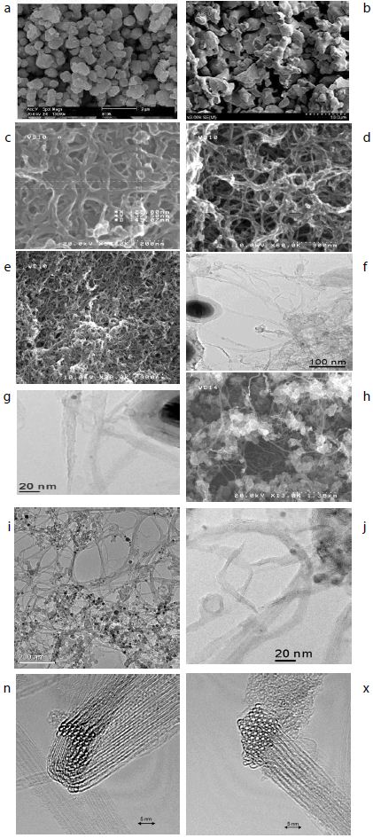
Figure 1: a-Ti powder morphology, b- Nb powder morphology, c, d, e, h- SEM of SWCNTs morphologies, f, g, j, k- TEM of SWCNTs with Fe catalyst, n, x: Bundles of SWCNTs.

Mechanical Alloying

The equilibrium phase diagram of NbTi binary alloy system. The phase diagram shows that the β-phase allotropic phase transformation, for pure titanium, occurs at 1155 K. Since niobium acts as a strong a-phase phase stabilizer, niobium additions result in a significant lowering of at β-type phase transformation temperature. It can be seen that Nb-Ti binary alloys with more than 50 wt% Nb can retain the β-phase at very low temperatures. Therefore, to obtain β-type phase stabilized Nb-Ti alloy, as received Ti and elemental Nb were blended together according to the desired optimum stoichiometric composition, i.e. 50 wt% titanium and 50 wt% Nb, NbxTi (x=50 at%) (further referred as Nb50Ti). The Nb and Ti powders were mechanically alloyed under argon atmosphere at room temperature, using planetary ball mill with stainless steel balls and vials-operating at a rotation speed of 200 rpm. To obtain homogenized and fine powder mixtures, the powder mixtures of NbxTi (x=50 at%) were ball-milled at a high speed of 200 RPM for 10 to 20 by using WC balls (diameter: 3 mm) and ethanol as the milling media. The ball to powder ratio was kept 6:1. To understand the microstructural evolution during ball-milling process, the mixture powder Nb and Ti samples were analyzed after mechanical milling time of 10 and 20 hours and then the powders mixture containing Nb50Ti were mixed with addition of 0.4 to 0.8 wt% of SWCNTs after preliminary treatment of SWCNTs was carried out to minimize the agglomerate of the added SWCNTs. Firstly; the weighed SWCNTs were immersed into acetone for about 5 h, and then were ultrasonically dispersed for 4 h. Secondly, the treated SWCNTs (0.4 to 0.8 wt% mixed with the former ball-milled blend Nb50Ti by magnetic agitation for 8 h and then ball milled for 10 h. The ball-milled intermetallic nanocomposites were prepared by wet milling in anhydrous alcohol for 3 h.

Finally, the powder mixtures with dispersed SWCNTs were dried by rotary evaporator under vacuum condition and were sieved to 70 mesh [49,50].

Consolidation of Powders via Spark Plasma Sintering

The field assisted sintering Technics-Spark Plasma Sintering (FAST-SPS) method is an effective technique for the compaction of powder materials. A main characteristic of this method is the direct heating of the pressing tool and/or the sample by pulsed direct electrical current with low voltage. This results in high heating rates and allows for short treatment times in order to obtain highly compacted sinter bodies. The material transport (e.g. by diffusion) occurring during the sintering process can also be used for performing chemical reactions. Especially the conditions during the FAST-SPS process allow the use of the method also as an alternative synthesis route for intermetallic compounds, of which, some can be obtained only with difficulties by other techniques.

The intermetallic matrix nanocomposites ball-milled powders were consolidated via FAST-SPS-FCT method under high vacuum conditions using graphite die and punch at an applied external pressure of 60 MPa. In order to prepare high density compacts with negligible porosity, temperature and compression pressure was programmed to rise simultaneously, until stabilized to the maximum temperature and compression pressure, followed by holding the system at the same sintering conditions for 1.6 ks. Circular compacts with diameter 20 mm and thickness 4 mm were prepared using FAST-SPS-FCT. The sintering conditions used in this study are provided in Table 1. The tree sintering schedules are. In the FAST-SPS-FCT, machine temperature profile, punch displacement or shrinkage and displacement velocity are presented in the FAST-SPS FCT machine (Table 1 and Figure 2).

Table 1: FAST-SPS synthesis parameters

Sintred Samples

T (°C)

Time of the cycle (mn)

Heating rate (°C/min)

P (MPa)

Ar (Sccm)

Wt.% SWCNTs d=1 nm

Current (A)

β-type phase Nb50Ti

1200

10

100

70

200

00

3000

β-type phase Nb50Ti/0.4 Wt% SWCNTs

1200

10

100

70

200

0.4

3000

β-type phase Nb50Ti/0.8 Wt% SWCNTs

1200

10

100

70

200

0.8

3000

fig 2

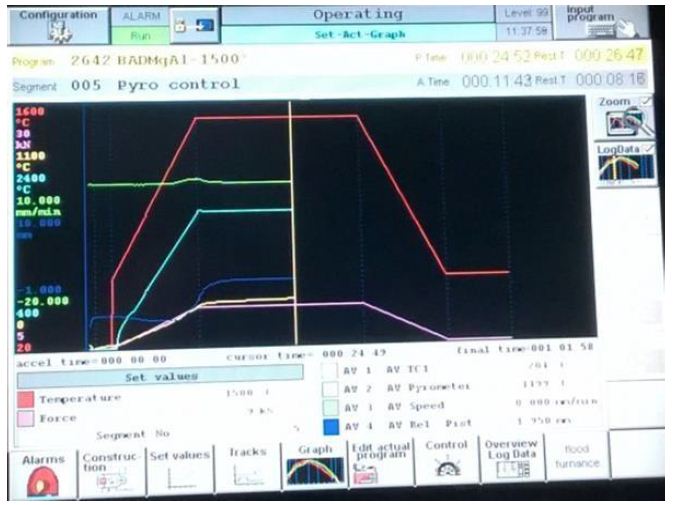
Figure 2: Variation of die displacement or shrinkage, temperature and applied pressure in dependence on the heating time during the FAST-SPS the sintered samples.

The resulting samples β-type phase Nb50Ti and β-type phase Nb50Ti/0.4-0.8 Wt% SWCNTs ultrafine powder mixtures were sintered by FAST-SPS in graphite dies (inner diameter of 20 mm) coated with graphite sheet lubricant atomized with cBN at 1273 to 1473°K in vacuum. Table 1 presented the sintering condition of intermetallic Nb50Ti and intermetallic nanocomposite Nb50Ti/SWCNTs 0.4-0.8. The applied pressure of 64 MPa was adjusted to the powder at room temperature and kept constant throughout the hot pressing process. The pressure was applied at the beginning of the sintering process because high green density is favorable for better densification rate by reducing the pores prior to the densification during heating. The heating rate was about 10°C/min and the dwelling time at terminal temperature was 60 min. The temperature was measured by an infrared pyrometer through a hole opened in the graphite die. Furthermore, for monitoring densification process, the shrinkage of the powder compact was measured by a displacement sensor during the sintering.

The dimensions of the finally sintered samples were about 20 mm in diameter and 3 mm in thickness after calculation of their weight using the densities values (Figure 3).

The mixtures were loosely compacted into a graphite die of 20 mm in diameter and sintered in the vacuum (1 Pa) at various temperatures using an FAST-SPS-FCT apparatus at the sinter Labs:

  1. ENSICAEN, 6, Boulevard Maréchal Juin, CS 45053 14050 Caen Cedex 04,
  2. Dipartimento di Ingegneria Chimica e Materiali, Universita` di Cagliari, Piazza d’Armi, 09123, Cagliari,
  3. Tycho lab, Institut fur Physik, universitat Rostock Germany FAST-SPS-1050, Sumitomo Coal Mining Co. , Germany). A constant heating rate of 120°C/min was employed, while the applied pressure was 65 MPa. The on/off time ratio of the pulsed current was set to 10/2 in each run. The maximum current reached approximately 3000 A during sintering.

The soaking time at high temperatures was within 10 min. The upper ram of the FAST-SPS apparatus was fixed, while the displacement of the shifting lower press ram was recorded in order to analyze the synthesis and sintering. The sintered samples are presented in the Figure 3.

Density of the sintered samples was measured by the Archimedes’ using the densimeter type Micromiritics Accupyc 1330.

fig 3

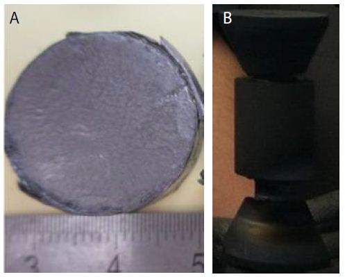
Figure 3: (a) Sintered samples in the die diameter of 20 mm of Nb50Ti /SWCNTs powder intermetallic nanocomposite. (b) Punch, die and spacer with 10 mm of diameter.

Mechanical Properties

Mechanical properties of the sintered compacts were evaluated the microhardness at the top were measured by a diamond Vickers hardness tester (MVK-H1, Meter-Mitutoyo, Japan).

The indentation loads, ranging from 2 to 500 N, were applied for 15 s for each measurement. The fracture toughness was measured using the Vickers indentation by the measurement of the producing failler.

In this study, 06 measurements for each sintering sample process were fabricated to obtain an average relative density and microhardness.

Young’s modulus of the composites was determined by ultrasonic wave transition method measuring the velocity of ultrasonicsound waves passing through the material using an ultrasonic flaw detector (Panametrics Epoch III). The microhardness and the fracture toughness were determined by the Vickers indentation method applying load of 294 N (HV30) and 490 N (HV50), by a Future Tech FLC-50VX hardness tester. For each sample, 6 indentations were made and the stress intensity factor KIC was calculated from the length of Palmqvistcracks which developed during a Vickers indentation test using E. Rocha-Rangel’s equation.

The wear resistance and the friction coefficient (tribological behavior will be performed in the near future. The hardness (H) and the toughness (KIC) of the manufactured samples were measured under ambient conditions using the instrumented Vickers indentation method (ZwickRoell, ZHU 2.5 apparatus). The impression diagonal (2a) was measured, and the hardness values were calculated according to the following relation:

Hv=(1.8544*F)/(2a)2                                                        (1)

The fracture toughness was also calculated by indentation fracture (IF) method according to the equation:

KIC=0.16Hva1/2(c/a)-3/2                                                    (2)

Where Hv was the Vickers hardness, a was the half-length of the indentation diagonal and c was the half-length of the median crack generated by indentation. Generally, the fracture toughness measured by IF method were fluctuating values with relatively large deviations due to the phase distribution and measurement errors of calculation. Thus a linear regression model was applied to get a reliable value of indentation fracture toughness.

To obtain the values of A, B and R2, a series of indentation loads (10 N, 50 N, 100 N, 300, 500 N) were applied to get the relations of P and c3/2

Where P was the indentation load. Through the combination of equations (1) and (2), the linear relation between P and c3/2was obtained:

P=Ac3/2 + B                                                           (A=KIC/0.075) (3)

A linear regression analysis was applied to the relations of P and c3/2 by the least square method. Where A was the slope, B was the intercept. To obtain the values of A and B.

In addition, a high determination coefficient (R2) was obtained through the linear regression model. Hence, when combined with the linear regression model, IF was shown to be an effective method in the evaluation of fracture toughness for its convenience and material saving. The compressive strength and the compressive modulus of elasticity of the produced alloy and nanocomposite were determined from compression tests carried out on the cylinders, at room temperature, at a strain rate of 10-3·s-1.

In vitro biocompatibility experiments to be performed using human bone marrow stromal cells (hBMSC). For isolation of hBMSC, bone marrow aspirates will be collected from bone marrow donors (age: 32 ± 3 years) at the CHU-Bone Marrow Transplantation Centre Hospital ibn Rochd-Annaba-Algeria to the laboratory.

The study will be approved by the local ethics commission. The donors will be informed and gave their approval. hBMSC were isolated using the method described by.

Bone marrow aspirate will be diluted 1:5 with 0.5% human serum albumin (HSA) in phosphate-buffered saline (PBS) and applied to a Percoll density gradient (d=1.073 g/mL). After centrifugation at 900 g for 30 min at 25°C, mononuclear cells in the interface will be harvested and filtered through a nylon cell strainer. The cells will be re-suspended in DMEM containing 10% heat-inactivated fetal calf serum FCS and antibiotics. After 24 h, non-adherent cells were removed. When the adherent cells reached about 90% confluence, they were trypsinized with 0.05% trypsin/0.02% EDTA (v/v) in PBS and sub-cultured. For the experiments, 5000 hMSC/cm2 were deposited on the test specimen. Metabolic activity was determined by the MTS assay (Cell Titer 96 Aqueous One Solution Proliferation Assay) 24 h after plating. Conditioned medium was replaced by fresh medium containing 10% of MTS dye solution. After 2 h of incubation at 37°C in a humidified CO2 incubator, 80 mL cellular medium was transferred to a 96-well plate and the absorbance of the formed MTS formazan dye will be measured photometrically at 490 or 655 nm.

In terms of statistics, the results of the quantitative parameters are presented as mean ± standard deviation (together with median and variance, in some cases). Significance of difference of the pT, ultimate compression strength, and compressive modulus of elasticity results between the two study groups was performed using the Mann-Whitney test, with significance denoted at p < 0.05. The superconducting measurement of critical temperature, critical current density and critical magnetic field on the nanocomposites samples for the superconducting magnet in ITER apparatus.

Results and Discussion

Phase and Microstructural Analysis

The general phase analysis was carried out at ambient temperature by XRD analysis with a CuKα (λ=1.5406 Å) source at 40 kV voltage and 30 mA current. The microstructural characterization of the initial powders, ball-milled powders, and consolidated compacts was carried out by FESEM equipped with back scattered electron (BSE) and electron back scattered diffraction (EBSD) facilities. Primary chemical characterization was carried out by Energy Dispersive X-ray Spectroscopy (EDS) technique. The diameter of single consists of β-type phase Nb50Ti about 24 ± 2 µm. The XRD pattern taken from the cross-section of the sintered samples is shown in Figure 4. Both a main β-phase and a minor α-phase are identifiable. The amount of α-phase was evaluated as 10 ± 2% by the Le Bail method. The appearance of α-phase can be mainly attributed to a slow cooling rate as the samples had to be cooled inside of FAST-SPS-FCT chamber. After slow cooling, the presence of the ω phase precipitates (hexagonal structure, space group P6/mmm) is also possible because, according to the literature data ω-phase precipitates may form during a slow quenching from the β-type phase region or during isothermal aging. The presence of the ω-phase is hard to detect from the XRD patterns but it was confirmed by TEM studies in a previous work [51-53].

The XRD patterns of the pre-mixed and ball-milled Nb50Ti powder mixtures (ball-milled from 10 to 20 haours) are shown in Figure 4a. These XRD patterns were compared with the JCPDS data of the oxides of Ti and Nb. No visible oxide peaks were found to be present in the XRD patterns of the ball-milled powders, indicating the absence of any substantial quantity of oxides. However, an analysis of the XRD profiles clearly shows a broadening of peaks with increasing milling time from 10 to 20 h. Sample made by FAST-SPS-FCT of intermetallic nanocomposite β-type phase Nb50Ti/0.4-0.8 Wt% SWCNTs ball-milled powder. At the Figure 4 is represented the higher resolution of its (101) peak.

fig 4

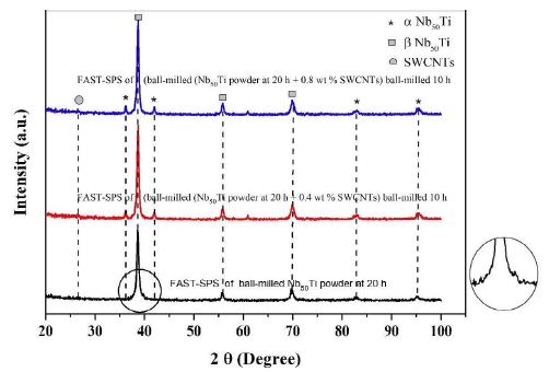
Figure 4: X-ray diffractometer (XRD), patterns of blue line, FAST-SPS of (ball-milled (Nb50Ti powder at 20 h + 0.8 wt% SWCNTs) ball-milled 10 h. Red line, FAST-SPS of (ball-milled (Nb50Ti powder at 20 h + 0.4 wt% SWCNTs) ball-milled 10 h. Black line FAST-SPS of ball-milled Nb50Ti powder at 20 h. A higher resolution of its (101) peak.

 
fig 4a

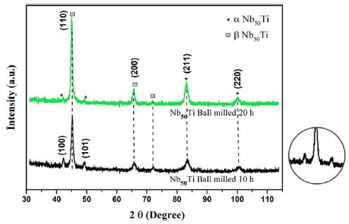
Figure 4a: X-ray diffractometer (XRD) patterns of green line Nb50Ti ball-milled 20 h. Black line Nb50Ti ball-milled 10h. A higher resolution of its (101) peak.

Figure 4a shows the XRD profiles of the ball-milled powders compacts sintered at 1473°K. These patterns depict and illustrate the microstructural/phase evolution at the sintering temperature. It can be clearly observed that the sintering temperatures led to almost formation of various new alloy phases such as Ti-rich α-type phase, and β-type phase. In particular, α, β-type phase, and pure niobium were formed at lower temperatures.

Ball-milled powder was sieved to select particle sizes of 20 ± 2 µm and its flowability was determined to be very good. Therefore, the ball-milling produced powder was found to be suitable for use in FAST-SPS-FCT. The broadening of the peaks in the XRD spectra at different stages of the ball-milling process suggests the formation of an ultrafine grain structure and an accumulation of strain in the lattice, which is typical for ball-milling related to severe mechanical deformation [54]. The ball-milling process, the morphology and phase composition of the powder were discussed in detail in the previous work of the authors (Figures 4 and 4a).

Structural and Morphological Characterization

To acquire the information on the distribution of various phases in the sintered samples, Energy-Dispersive X-ray Spectroscopy (EDS) analysis was performed on the samples. Although it is not possible to identify the phases by EDS directly, it is still very useful to detect elemental distribution of Nb, Ti and SWCNTs in samples. Thus, EDS analysis together with XRD results can provide more information regarding nature of FAST-SPS of compact powder.

The FESEM images and their corresponding elemental distribution maps of previous ball-milled and then FAST-SPS powders at T=1473°K are shown in Figure 5a. Figure 5b shows the morphology, and their corresponding elemental distribution, of the powder mixture (Nb, Ti, SWCNTs).

Zhang et al. [55] has reported that cold welding and severe plastic deformation is the dominant mechanism which operates during mechanical milling of ductile Nb powder particles. Due to severe plastic deformation and cold-welding, Nb particles become hard and brittle, leading to their fragmentation during subsequent mechanical milling. As a result, relatively coarser Nb particles are obtained in the ball-milled powder mixture. It would also be worth mentioning that such a deformation and fracturing of particles leads to the formation of niobium particles with energetically activated surface.FESEM images and their corresponding elemental distribution maps phase evolved as the major phase in the sintered compacts and the intensity of the peaks corresponding to the β-type phase increased with increasing of amount of SWCNTs at the sintering temperature. Finally, β-type phase appeared to be the major phase formed in the compacts sintered at 1473°K. They consisted of primarily beta phase together with extremely small amounts of alpha phase. Microstructure of the Nb50Ti alloy intermetallic ball-milled 20 h compacts sintered at T=1473°K is illustrated in Figure 5a through typical FESEM micrographs collected in back scattered electron (BSE) mode. The sintered Nb50Ti and intermetallic nanocomposite Nb50Ti/SWCNTs samples exhibited very high density, approximately 94%, with appearance of minimum porosity. The microstructure of low-temperature sintered (1473°K) Nb40 Ti intermatallic alloy compacts (Figure 5a) show three different color composition: (I) bright colored phase, which is the pure Nb phase as verified by energy dispersive X-ray spectroscopy (EDS) analysis and contains Nb and Ti; (II) grey-colored phase, which is the β-type phase and contains Nb50Ti; and(III) comparatively dark-colored phase, which is the α-phase and contains Nb50Ti. Similarly, the microstructure of the Nb50Ti/0.8 wt% WCNTs intermatallic nanocomposite compacts sintered at moderate temperature (1473°K), as shown in Figure 5b, also demonstrated three different phases similar to the ones observed in the low temperature sintered Nb50Ti compacts. It can be observed that, the fraction of the bright and the dark phases decreased whereas the fraction of the grey phase increased in the matrix with increasing of amounts of SWCNTs from0.4 wt% to 0.8 wt%, which could be attributed to the increased inter-diffusion of Ti and Nb and SWCNTs at the sintering temperature. The EDS analysis clearly demonstrated that the grey phase corresponds to the β type phase composition. Therefore, the above results clearly show that the lower sintering temperature result β type phase rich Nb50Ti intermetallic alloy with increased homogeneity. Clearly, these results are in conformity with the phase evolution as observed via XRD results, as already shown in Figure 4.

The FESEM represented separately each α-phase and β-type phase. For Nb50Ti compacts sintered at 1473°K, the calculated average grain size for α and β-type phase was 2.9 ± 1.02 μm and 9.61 ± 4.20 μm, respectively (Figures 5a and 5b).

fig 5

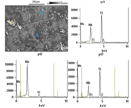
Figure 5: High magnification micro structural representative FESEM image and EDS spectra obtained on tree different regions of the surface of the sample made of sample of Nb50Ti ball- milled 20 h and sintered at T=1473°K.

fig 5a

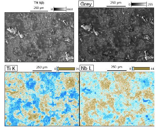
Figure 5a: High magnification micro structural representative FESEM image their corresponding elemental distribution maps (Nb, Ti) of previous sample of Nb50Ti ball-milled 20 h of polished and etched surface sintered at T=1473°K.

fig 5b

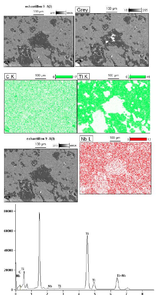
Figure 5b: High magnification micro structural representative FESEM image their corresponding elemental distribution maps (Nb, Ti and C) of FAST-SPS (ball-milled Nb50Ti powder at 20 h + 0.8 wt% SWCNTs) ball-milled 10 h of polished and etched surface sintered at T=1473°K. Its higher resolution global analysis EDS pics is presented.

Finally, the average grain size α-type phase and β-type phase of Nb50Ti/0.8 wt% SWCNTs intermatallic nanocomposite compacts sintered at 1473°K was 1.75 ± 0.4 μm and 4.75 ± 0.83 μm, respectively, in the Figure 6 is presented the homogenous distribution of carbon nanotubes (Figure 6).

fig 6

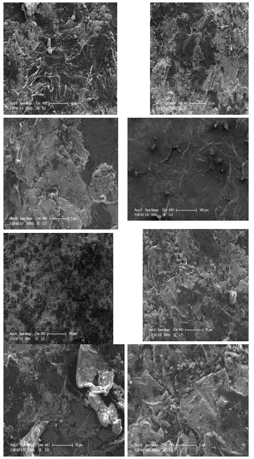
Figure 6: High magnification micro structural représentative FESEM of FAST-SPS (ball-milled Nb50Ti powder at 20 h + 0.8 wt% SWCNTs) ball-milled 10 h of polished and etched surface sintered at T=1473°K.

High magnification micro structural representative FESEM image sample of Nb50Ti ball-milled 20 h of polished and etched surface sintered at T=1473°K, showing the precipitates embedded in the β-type phase matrix intermetallic nanocomposites (Figure 7).

fig 7

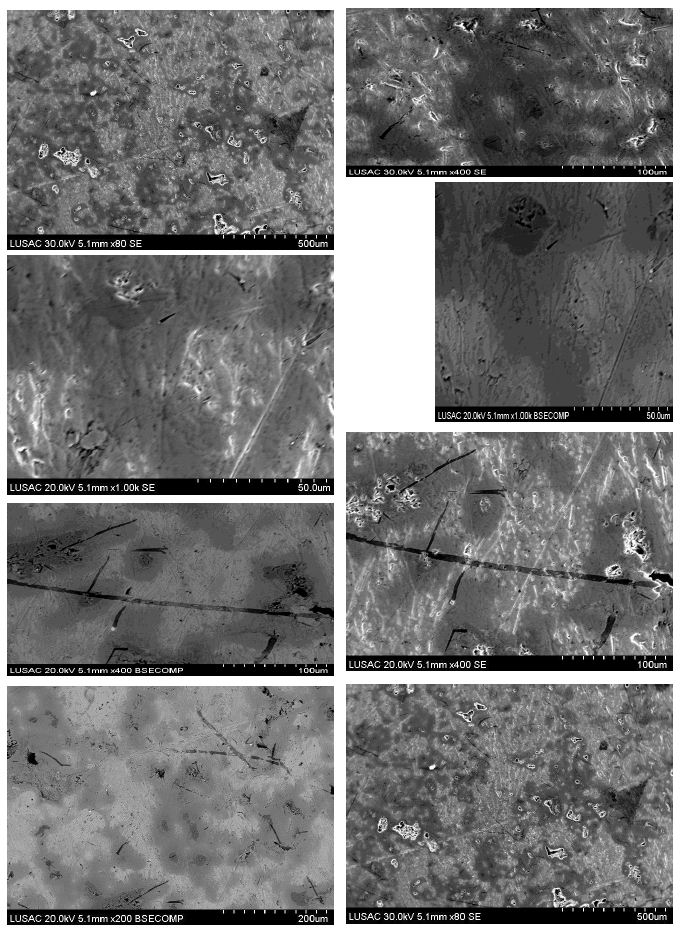
Figure 7: High magnification micro structural representative FESEM image sample of Nb50Ti ball-milled 20 h of polished and etched surface sintered at T=1473°K, showing the precipitates embedded in the β- type phase matrix.

These results clearly demonstrate that fine-grained β-type phase Nb50Ti intermetallic alloy and its nanocomposite based on SWCNTs can be successfully prepared via present processing technique FAST-SPS after sequentially ball-milling wherein the high yield of the material with good density and defined porosity remains ensured for biomedical and nanotechnologies applications by FAST-SPS method.

Mechanical Testing

Mechanical Properties

Density and Porosity of Sintered Samples

The theoretical density of the nanocomposite used for obtaining relative density was calculated using a rule of mixture, using the densities of two constituent phase (Nb50Ti=6 5.7 g/cm3, ρSWCNTs=2.25 g/cm3) with the given FAST-SPS processing parameters, the Nb50Ti without addition of 0.4 to 0.8 wt% SWCNTs sample exhibited best densification with relative density greater than 94.5%, with the similar processing parameters with addition of SWCNTs. The relative density increases with the addition of SWCNTs. The Nb50Ti with addition of 0.4 to 0.8 wt% SWCNTs intermetallic nanocomposites at T=1200°C exhibited relative density of about 97.2%. Depending on the final density to be achieved, the FASY-SPS operating condition were properly chosen, that is, 1200°C, 75 MPa for 10 min, to obtain a highest relative density for the intermetallic nanocomposites for 7, 23, and 2.8% compacts porosity, 94.5, 97.06 and 97.6, respectively.

The porosity (pT) for the FAST-SPS fabricated cylinders was 7% ± 1% (median=7.0%; variance: 0.4%), which is in good agreement with the value obtained using density determined by simply dividing the mass of the cylinder by its volume for Nb50Ti for biomedical application. The porosity is mainly formed by spherical pores, which result from the gas uptake by the powder during the ball milling process. These spherical pores and such a relatively low porosity are typical for alloys fabricated using FAST-SPS.

For FAST-SPS cylinders, the size of the micropores and pT were 3–12 µm and 3 -2.6% ± 0.4% (median=3.0%; variance=0.06%), for Nb50Ti with addition of 0.4 to 0.8 wt% SWCNTs which is in good agreement for biomedical application (Table 2 and Figure 8).

Table 2: Basic physical–mechanical properties of the samples obtained by ball-millig and then FAST-SPS

Sintred samples

β-type phase

Nb50Ti

β-type phase

Nb50Ti/0.4 Wt% SWCNTs

β-type phase

Nb50Ti/0.8 Wt% SWCNTs

Relative density (g/cm3) (%)

94.04

97.06

97.6

Young’s modulus (relative Young’s) (GPa)

60

62

64

Compressive elastic straingh (MPa)

620

630

650

Compressive elastic modulus (MPa)

124

126

130

Porosity (pT) (%)

7

3.0

2.8

fig 8

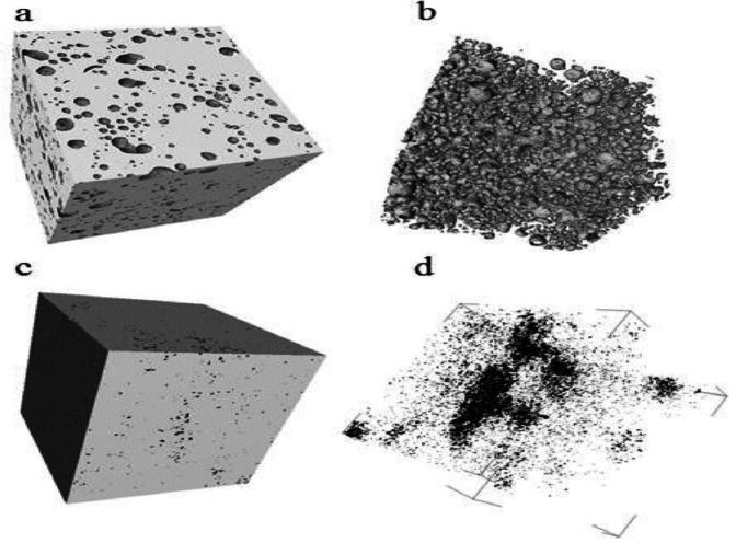
Figure 8: Micro-computed tomography (µCT) images of (a) sample manufactured by FAST’SPS of (ball-milled (Nb50Ti powder at 20 h + 0.8 wt% SWCNTs) ball-milled 10 h.(b) its inner porous architecture (from image analysis); (c) sample made FAST-SPS of ball- milled Nb50Ti powder at 20 h.

Vickers Microhardness of Sintered Samples

Hardness values were measured on the bulk Nb50Ti alloy and its nanocomposite based SWCNTs compact samples sintered at T=1473°K. The variation in the hardness with the amounts of SWCNTs at sintering temperature is shown in Figure 9. It can be seen that the average hardness increases significantly with increase in the amounts of SWCNTs from 0.4 to 0.8.

fig 9

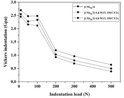
Figure 9: Representation of the variation of Vickers micro hardness function of the indentation leads of the sintered samples using a 20-mm.

Nevertheless, it would be worth noticing that the hardness of the specimens with coarser grain size and higher amounts of softer β-type phase demonstrates higher average hardness values as compared to those having relatively harder α phase. Interestingly, it can also be observed that the standard deviation decreased from 2.35 to 2.65 Gpa (Table 2) with increasing of amounts of SWCNTs. These results can be explained with respect to the volume fraction of various phases and homogeneity of microstructures. The average hardness of any specimen is essentially an average of the hardness of these individual phases coupled with volume fractions resulting in better mechanical properties and ductility.

According to the above results, it can be concluded that the Vickers hardness has been improved by adding SWNCs and enhanced with the fracture toughness value giving a better ductility and defined porosity for the reinforced β-type phase samples for bio-application. With this addition in the matrix, the electro discharge among powders may lead to self-heating and purification of the particle surface, resulting in activation of the formation of the nanocomposites. The addition of SWNCs plays an important binder less role (the ductility) in the propagation of failler in this nanocomposites and thus enhance the fracture toughness in comparison with his higher hardness.

Fracture Toughness (KIC) of Sintered Samples

Niobium is known by his ductility and superconductivity. In this context we proceeded to the determination of the tenacity of the sintered samples as well as the determination of the parameters associated with this calculation. But unfortunately we could not determine the critical cracking load due to ductility of the sintered samples.

Vibronic Properties

Raman Spectroscopy Analysis

To verify the XRD experimental results Raman spectroscopy is a powerful technique for characterizing degree of disorder of CNTs. SWNTs with a Raman spectroscopy measurement of 513.5 nm were studied. In general, Raman spectroscopy can observe the distribution of dielectric bonding SP3, D band and metallic bonding SP2, G band [55-61].

Different regions of the sample. In Figure 10 is presented which depicts the absence of radial breathing mode (RBM), which is attributed to the absence of SWCNTs. The first-order Raman spectra of MWCNTs has a small peak at 1364 cm-1 and strong peak at 1590 cm-1. A band at 1364 cm-1 (D) arises due to the presence of disorder in the carbon system as well as breathing of hexagons at border of crystalline areas of nanoparticles and fine crystallite. A sharp and narrow high-intensity band occurred at 1590 cm-1 (G band) which approved high crystallinity of graphite and graphitic structure of graphene. 2D or G-band peak at 2705 cm-1 and at 2720 cm-1 for second order of D band. They investigated that 2D band peak of synthesized graphene have no shoulder, which corroborates the characteristics of graphene with few layers which is a characteristic of graphene with more layer and ascribed to the overtone of first-order D band (1364) cm-1.

fig 10

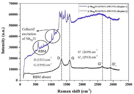
Figure 10: Raman spectra of FAST-SPS (ball-milled Nb50Ti powder at 20 h+0.8wt% SWCNTs) ball-milled 10 h of polished and etched surface sintered at T=1473°K (region 1 and 2).

The ratio of D band and G band demonstrates the degree of disorder induced in bulk samples. If both bands have equal intensity, then there is a high degree of disorder in the bulk samples. The relations among D band, G band and RBM play predominant role to distinguish between different variants of CNTs.

The experimental results are shown in Figure 10. The peak positions of SWCNTs were similar, but the ratios of the D band 1380 cm-1 and the G band 1590 cm-1 were quite different and collective excitation of β-type phase from the difference in the ratios, it was found that region 1 have higher dielectric properties than region 2 (Figure 10) because SWCNTs have underwent a transformation during heating of sintering to DWCNTs or MWCNTs in high vacuum.

To analyze these experimental results qualitatively, we modeled the composite system which is composed of CNTs nanoparticles in Nb50Ti host medium. The CNTs have both metallic and semiconducting properties (region 3 and 4) (Figure 10a) because they consist of diverse nanotubes and contact junctions [62,63].

fig 10a

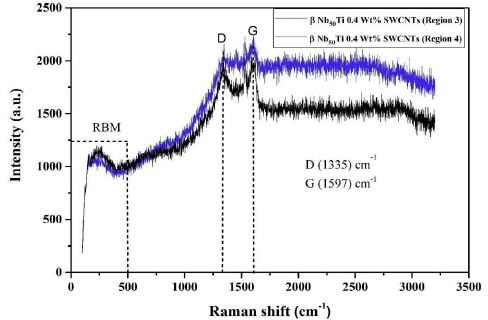
Figure 10a: Raman spectra of FAST-SPS (ball-milled Nb50Ti powder at 20h + 0.8wt% SWCNTs) ball-milled 10 h of polished and etched surface sintered at T=1473°K (region 3 and 4).

Describing these properties, the dielectric function of CNTs (ϵCNTs) CNTs can be represented as a combination of the Drude and Lorentz harmonic oscillator models [64] (Figures 10 and 11).

fig 11

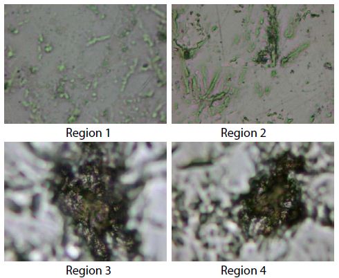
Figure 11: Raman images caption, region 1 and 2 corresponding to carbon nanotubes and β-type phase Nb50Ti. The region 3 and 4 correspond to carbon nanotubes.

Conclusions

vIn this study, intermetallic alloys fine-grained β-type phase matrix Nb50Ti/SWCNTs nanocomposite was successfully prepared by ball-milling of titanium elemental niobium and SWCNTs powders followed by spark plasma sintering. The final ball-milling (Nb50Ti/SWCNTs) resulted in a very high yield of milled powders, the powder had micron-sized Nb50Ti and a nano-sized (Nb50Ti/SWCNTs).

After the FAST-SPS the obtained material was a small amount of α and major β-type phase. The microstructure and the phase evolution in the sintered Nb50Ti compacts depend on the SWCNTs addition because now diffusion between Nb, Ti and SWCNTs was not observed, and almost complete β-type phase was achieved after sintering at 1473 K. Spark plasma sintering demonstrated the capability to yield fine grained microstructure even after sintering at low temperatures. The grain size of the synthesized Nb50Ti strongly depends on the on the SWCNTs addition, i.e. the grain size of α and β-type phases increases with of amounts of SWCNTs.

Compressive strength and modulus of the FAST-SPS -manufactured β-type phase were 620 ± 6 MPa and 124 ± 2 GPa, respectively, whereas the corresponding values for the intermetallic nanocomposite β-type phase matrix were 650 ± 8 MPa and 130 ± 2 GPa. For each of these properties, the difference in results for the two materials may be attributed to the difference in phase composition and pT.

These findings suggest that the combination of ball-milled of Nb50Ti/SWCNTs powder and FAST-SPS may have promise for use in manufacturing load-bearing metallic orthopaedic components, such as the femoral stem of a hip implant and in superconductor magnets (Poloidal Field Coils) in ITER.

The average hardness increased significantly with increasing of SWCNTs wt% at the sintering temperature 1473 K. The increasing average hardness with increasing of SWCNTs wt% at the sintering temperatures of 1473 K at 0.4 to 0.8 wt% of SWCNTs was attributed to the increasing volume fraction of β-type phase together with the embrittlement of β-type phase due to SWCNTs entrapment in the sintered compacts. In other hand SWCNTs increase the ductility and then the fracture toughness by stopping failler of the materials. The superconducting and wear properties will be performed in the near future.

Acknowledgments

The present work was supported by the Algerian Ministerium of Higher Education and Scientific Research and the PRFU project under contract N° A11N01UN240120220005. These supports are gratefully appreciated.

Conflicts of Interest

The authors declare no conflict of interest.

Declaration of Interests

  • The authors declare that they have no known competing financial interests or personal relationships that could have appeared to influence the work reported in this paper.
  • The authors declare the following financial interests/personal relationships which may be considered as potential competing interests.

References

  1. Attar H, Löber L, Funk A, Calin M, Zhang LC, et al. (2015) Mechanical behavior of porous commercially pure Ti and Ti–TiB composite materials manufactured by selective laser Materials Science and Engineering: A 625: 350-356.
  2. Bönisch M, Stoica M, Calin M (2020) Routes to control diffusive pathways and thermal expansion in Ti-alloys. Scientific Reports 10: 1-9. [crossref]
  3. Attar H, Calin M, Zhang LC, Scudino S, Eckert J (2014) Manufacture by selective laser melting and mechanical behavior of commercially pure titanium. Materials Science and Engineering: A 593: 170-177.
  4. Attar H, Prashanth KG, Chaubey AK, Calin M, Zhang LC, et al. (2015) Comparison of wear properties of commercially pure titanium prepared by selective laser melting and casting processes. Materials Letters 142: 38-41.
  5. Zhang LC, Attar H, Calin M, Eckert J (2016) Review on manufacture by selective laser melting and properties of titanium based materials for biomedical Materials Technology.31: 66-76.
  6. Lekka CE, Gutiérrez-Moreno JJ, Calin M (2017) Electronic origin and structural instabilities of Ti-based alloys suitable for orthopaedic Journal of Physics and Chemistry of Solids 102: 49-61.
  7. Bönisch M, Calin M, Van Humbeeck J, Skrotzki W, Eckert J (2015) Factors influencing the elastic moduli, reversible strains and hysteresis loops in martensitic Ti–Nb alloys. Materials Science and Engineering: C 48: 511-520. [crossref]
  8. Okulov IV, Bönisch M, Okulov AV, Volegov AS, Attar H, et al. (2018) Phase formation, microstructure and deformation behavior of heavily alloyed TiNb-and TiV-based titanium Materials Science and Engineering: A 733: 80-86.
  9. Okulov IV, Volegov AS, Attar H, Bönisch M, Ehtemam-Haghighi S, et al. (2017) Composition optimization of low modulus and high- strength TiNb-based alloys for biomedical Journal of the Mechanical Behavior of Biomedical Materials 65: 866-871. [crossref]
  10. Panigrahi A, Bönisch M, Waitz T, Schafler E, Calin M, et al. (2015) Phase transformations and mechanical properties of biocompatible Ti– Z16.1 Nb processed by severe plastic deformation. Journal of Alloys and Compounds.628: 434-441.
  11. Prashanth KG, Zhuravleva K, Okulov I, Calin M, Eckert J, et al. (2016) Mechanical and corrosion behavior of new generation Ti-45Nb porous alloys implant devices. Technologies 4: 33.
  12. Gennari S, Maglia F, Anselmi-Tamburini U, Spinolo G (2003) SHS (Self-sustained high-temperature synthesis) of intermetallic compounds: effect of process parameters by computer simulation. Intermetallics 11: 1355-1359.
  13. Liu E, Jia J, Bai Y, Wang W, Gao Y (2014) Study on preparation and mechanical property of nanocrystalline NiAl intermetallic. Materials & Design 53: 596-601.
  14. Geist D, Gammer C, Rentenberger C, Karnthaler HP (2015) Sessile dislocations by reactions in NiAl severely deformed at room Journal of Alloys and Compounds.621: 371-377. [crossref]
  15. Liu E, Gao Y, Bai Y, Yi G, Wang W, et al. (2014) Tribological properties of self-lubricating NiAl/Mo-based composites containing AgVO3 Materials Characterization 97: 116-124.
  16. Zhang JF, Shen J, Shang Z, Feng ZR, Wang LS, et al. (2012) Microstructure and room temperature fracture toughness of directionally solidified NiAl–Mo eutectic in situ composites. Intermetallics 21: 18-25.
  17. Zhuravleva K, Bönisch M, Prashanth KG, Hempel U, Helth A, et al. (2013) Production of porous β-Type Ti–40Nb alloy for biomedical applications: Comparison of selective laser melting and hot pressing. Materials 6: 5700-5712. [crossref]
  18. Bocanegra-Bernal MH, Dominguez-Rios C, Echeberria J, Reyes-Rojas A, Garcia-Reyes A, et al. (2016) Spark plasma sintering of multi-, single/double-and single-walled carbon nanotube-reinforced alumina composites: is it justifiable the effort to reinforce them?. Ceramics International 42: 2054-2062.
  19. Moshksar MM, Mirzaee M (2004) Formation of NiAl intermetallic by gradual and explosive exothermic reaction mechanism during ball Intermetallics 12: 1361-1366.
  20. Suryanarayana C (2001) Mechanical alloying and milling. Progress in Materials Science 46: 1-184.
  21. Beyhaghi M, Kiani-Rashid AR, Kashefi M, Khaki JV, Jonsson S (2015) Effect of powder reactivity on fabrication and properties of NiAl/Al2O3 composite coated on cast iron using spark plasma sintering. Applied Surface Science 344: 1-8.
  22. Umanskii AP, Polyarus EN, Ukrainets MS, Kapitanchuk LM (2015) Structure and tribotechnical characteristics of NiAl–CrB2 composite materials and Powder Metallurgy and Metal Ceramics 54: 53-59.
  23. Singla D, Amulya K, Murtaza Q (2015) CNT reinforced aluminium matrix composite-a review. Materials Today: Proceedings 2: 2886-2895.
  24. Alizadeh A, Abdollahi A, Biukani H (2015) Creep behavior and wear resistance of Al 5083 based hybrid composites reinforced with carbon nanotubes (CNTs) and boron carbide (B4C) Journal of Alloys and Compounds 650: 783-793.
  25. Zhang M, Hou X, Wang J, Li M, Hu S, et al. (2014) Interweaved Si@ C/CNTs&CNFs composites as anode materials for Li-ion Journal of Alloys and Compounds 588: 206-211.
  26. Peng T, Chang I (2014) Mechanical alloying of multi-walled carbon nanotubes reinforced aluminum composite powder. Powder Technology 266: 7-15.
  27. Vishlaghi MB, Ataie A (2014) Investigation on solid solubility and physical properties of Cu–Fe/CNT nano-composite prepared via mechanical alloying route. Powder Technology 268: 102-109.
  28. Mazaheri M, Mari D, Hesabi ZR, Schaller R, Fantozzi G (2011) Multi-walled carbon nanotube/nanostructured zirconia composites: outstanding mechanical properties in a wide range of temperature. Composites Science and Technology.71: 939-945.
  29. Pang LX, Sun KN, Ren S, Sun C, Fan RH, et al. (2007) Fabrication and microstructure of Fe3Al matrix composite reinforced by carbon Materials Science and Engineering: A 447: 146-149.
  30. Schmidt R, Pilz S, Lindemann I, Damm C, Hufenbach J, et al. (2017) Powder metallurgical processing of low modulus β-type Ti- 45Nb to bulk and macro-porous compacts. Powder Technology 322: 393-401.
  31. Wang Q, Cui G, Chen H (2021) Effect of the Ta addition on densification and mechanical properties of NbTi alloys prepared by Journal of Alloys and Compounds 868: 159106.
  32. Helth A, Gostin PF, Oswald S, Wendrock H, Wolff U, et al. (2014) Chemical nanoroughening of Ti40Nb surfaces and its effect on human mesenchymal stromal cell response. Journal of Biomedical Materials Research Part B: Applied Biomaterials 102: 31-41. [crossref]
  33. Gostin PF, Helth A, Voss A, Sueptitz R, Calin M, et al. (2013) Surface treatment, corrosion behavior, and apatite‐ forming ability of Ti‐ 45Nb implant Journal of Biomedical Materials Research Part B: Applied Biomaterials 101: 269-278. [crossref]
  34. Zhuravleva K, Bönisch M, Scudino S, Calin M, Schultz L, et al. (2014) Phase transformations in ball-milled Ti–40Nb and Ti–45Nb powders upon quenching from the ß-phase Powder Technology 253: 166- 171.
  35. Kauschke V, Gebert A, Calin M, Eckert J, Scheich S, et al. (2018) Effects of new beta-type Ti-40Nb implant materials, brain-derived neurotrophic factor, acetylcholine and nicotine on human mesenchymal stem cells of osteoporotic and non-osteoporotic PLoS One 13: e0193468. [crossref].
  36. Attar H, Calin M, Zhang LC, Scudino S, Eckert J (2014) Manufacture by selective laser melting and mechanical behavior of commercially pure Materials Science and Engineering: A 593: 170-177.
  37. Helth A, Pilz S, Kirsten T, Giebeler L, Freudenberger J, et al. (2017) Effect of thermomechanical processing on the mechanical biofunctionality of a low modulus Ti-40Nb alloy. Journal of the Mechanical Behavior of Biomedical Materials 65: 137-150. [crossref]
  38. Schmidt R, Hoffmann V, Helth A, Gostin PF, Calin M, et al. (2016) Electrochemical deposition of hydroxyapatite on beta-Ti-40Nb. Surface and Coatings Technology 294: 186-193.
  39. Gostin PF, Helth A, Voss A, Sueptitz R, Calin M, et al. (2013) Surface treatment, corrosion behavior, and apatite‐ forming ability of Ti‐ 45Nb implant Journal of Biomedical Materials Research Part B: Applied Biomaterials 101: 269-278. [crossref]
  40. Zhuravleva K, Chivu A, Teresiak A, Scudino S, Calin M, et al. (2013) Porous low modulus Ti40Nb compacts with electrodeposited hydroxyapatite coating for biomedical applications. Materials Science and Engineering: C 33: 2280-2287.
  41. Abdi S, Oswald S, Gostin PF, Helth A, Sort J, et al. (2016) Designing new biocompatible glass‐ forming Ti75‐ xZr10NbxSi15 (x= 0, 15) alloys: corrosion, passivity, and apatite formation. Journal of Biomedical Materials Research Part B: Applied Biomaterials 104: 27-38.
  42. Hynowska A, Blanquer A, Pellicer E, Fornell J, Surinach S, et al. (2015) Nanostructured Ti‐ Zr‐ Pd‐ Si‐ (Nb) bulk metallic composites: Novel biocompatible materials with superior mechanical strength and elastic recovery. Journal of Biomedical Materials Research Part B: Applied Biomaterials.103: 1569-1579.
  43. Calin M, Helth A, Gutierrez Moreno JJ, Boenisch M, Brackmann V, et al. (2014) Elastic softening of β-type Ti–Nb alloys by indium (In) additions. Journal of the Mechanical Behavior of Biomedical Materials 39: 162-174. [crossref]
  44. Hynowska A, Pellicer E, Fornell J, Gonzalez S, van Steenberge N, et al. (2012) Nanostructured β-phase Ti–31.0 Fe–9.0 Sn and sub-μm structured Ti–39.3 Nb–13.3 Zr–10.7 Ta alloys for biomedical applications: Microstructure benefits on the mechanical and corrosion performances. Materials Science and Engineering: C 32: 2418-2425.
  45. Calin M, Gebert A, Ghinea AC, Gostin PF, Abdi S, et al. (2013) Designing biocompatible Ti-based metallic glasses for implant Materials Science and Engineering: C 33: 875-883.
  46. Gutierrez Moreno JJ, Boenisch M, Panagiotopoulos NT, Calin M, Papageorgiou DG, et al. (2017) Ab-initio and experimental study of phase stability of Ti-Nb Journal of Alloys and Compounds 696: 481-489.
  47. Bendjemil B, Borowiak-Palen E, Graff A, Pichler T, Knupfer M, et al. (2004) Elimination of metal catalyst and carbon-like impurities from single-wall carbon nanotube raw material. Applied Physics A 78: 311-314.
  48. Selbmann D, Bendjemil B, Leonhardt A, Pichler T, Täschner C, et al. (2008) A parametric study of the synthesis and purification of single- walled carbon nanotubes using the high-pressure carbon monoxide Applied Physics A 90: 637-643.
  49. Bendjemil B, Mouyane M, Noudem JG, Bernard J, Reboul JM, et al. (2018) Sintering Behavior and Properties of cBN/TiC/SWCNTs or NC Ceramics Matrix Nanocomposites (CMNC’s) by Field Actived Sparck Plasma Sinter.
  50. Bendjemil B, Noudem JG, Mouyane M, Bernard J, Guhel Y, et al. (2020) Study of the Nanocomposite Mo2C (1-x)-TiC (x)-SWCNTs by Field Actived Sparck Plasma Sintering Journal of Advances in Nanotechnology 1: 30-47.
  51. Le Bail A (1995) Modelling the silica glass structure by the Rietveld method. Journal of Non-Crystalline Solids 183: 39-42.
  52. Moffat DL, Kattner UR (1988) The stable and metastable Ti-Nb phase diagrams. Metallurgical Transactions A.19: 2389-2397.
  53. Zhuravleva K, Bönisch M, Scudino S, Calin M, Schultz L, et al. (2014) Phase transformations in ball-milled Ti–40Nb and Ti– 45Nb powders upon quenching from the ß-phase Powder Technology 253: 166-171.
  54. Zhuravleva K, Scudino S, Khoshkhoo MS, Gebert A, Calin M, et al. (2013) Mechanical Alloying of β‐ Type Ti–Nb for Biomedical Advanced Engineering Materials 15: 262-268.
  55. Zhang DZ, Qin ML, Zhang L, Qu XH (2012) Fabrication and characterization of nanocrystalline Nb–W–Mo–Zr alloy powder by ball milling. International Journal of Refractory Metals and Hard Materials 32: 45-50.
  56. Hempel U, Müller K, Preissler C, Noack C, Boxberger S, et al. (2016) Human bone marrow stromal cells: a reliable, challenging tool for in vitro osteogenesis and bone tissue engineering Stem Cells International 14. [crossref]
  57. Herzer R, Gebert A, Hempel U, Hebenstreit F, Oswald S, et al. (2021) Rolled‐ Up Metal Oxide Microscaffolds to Study Early Bone Formation at Single Cell Resolution. Small 17: 2005527. [crossref]
  58. Vishnu J, Calin M, Pilz S, Gebert A, Kaczmarek B, et al. (2020) Superhydrophilic nanostructured surfaces of beta Ti29Nb alloy for cardiovascular stent Surface and Coatings Technology 396: 125965.
  59. Charifoulline Z (2006) Residual resistivity ratio (RRR) measurements of LHC superconducting NbTi cable IEEE Transactions on Applied Superconductivity 16: 1188-1191.
  60. Fang L, Feng L, Huixian G, Chao C, Bo L, et al. (2015) Comparison and analysis of twist pitch length test methods for ITER Nb3Sn and NbTi Rare Metal Materials and Engineering 44: 2095-2099.
  61. Dresselhaus MS, Dresselhaus G, Avouris P (2001) Carbon Nanotubes Synthesis, Structure, Properties, andApplications.
  62. Ugawa A, Rinzler AG, Tanner DB (1999) Far-infrared gaps in single- wall carbon Physical Review B 60: R11305.
  63. Hilt O, Brom HB, Ahlskog M (2000) Localized and delocalized charge transport in single-wall carbon-nanotube Physical Review B 61: R5129.
  64. Ahn JS, Kim KH, Noh TW, Riu DH, Boo KH, et al. (1995) Effective-medium theories for spheroidal particles randomly oriented on a plane: Application to the optical properties of a SiC whisker-Al2O3 composite. Physical Review B 52: 15244. [crossref]
fig 4

Effects of Photoenhanced Thin Oil Sheens on Survival and Growth of Newly Hatched Fishes: Sheepshead Minnow (Cyprinodon variegatus), spotted seatrout (Cynoscion nebulosus), and Red Drum (Sciaenops ocellatus)

DOI: 10.31038/AFS.2022444

Abstract

The release of polycyclic aromatic hydrocarbons (PAHs), related to oil spills, can have devasting effects on the environment. In the presence of ultraviolet (UV) light, PAHs can be photoenhanced into more toxic compounds, leading to increased toxicity in aquatic organisms as measured by 24-h survival. PAHs cause a suite of physiological consequences particularly in the early stages of fish development due to increased surface area to volume ratios and nascent immune systems. This study compared the impacts of photoenhanced thin oil sheens on the survival and growth of larvae of three ecologically important coastal fish species: sheepshead minnows (Cyprinodon variegatus), red drum (Sciaenops ocellatus) and spotted seatrout (Cynoscion nebulosus). PAH + UV exposure increased the toxicity of thin oil sheens in all three species, with red drum demonstrating the highest sensitivity. An acute oil exposure at 1-2 days post hatch (dph) increased the rate of latent mortality and oxidative stress in sheepshead minnows. Non-photoenhanced oil caused a significant decrease in the growth of 1-2 dph sheepshead minnows and spotted seatrout. Results from this study reveal long-term effects of oil exposure on fish growth and survival, which could lead to better restoration and conservation outcomes for these vital estuarine species.

Introduction

Petroleum can enter waterways through large spills such as the April 2010 Deepwater Horizon (DWH) oil spill and the March 1989 Exxon Valdez shipping container oil spill [1]. More frequently, however, oil is released on smaller scales through industrial discharge, smaller vessel spills, runoff from impervious surfaces, and commercial port usage [2]. Oil pollution can cause physical and chemical changes to the coastal environment, as well as negatively impact the health of marine organisms [3].

Oil contains polycyclic aromatic hydrocarbons (PAHs), which are a group of organic molecules that are composed of fused benzene rings which allow them to be lipophilic, readily bioaccumulated, and have mutagenic and carcinogenic properties [4-7]. The effects of PAHs, especially those found in crude oil, on marine organisms have been extensively researched and the documented effects are widespread across a wide range of organisms [8-26]. Effects such as deformities, reduced growth and reproduction, genetic and behavioral changes, and altered physiological functions have been seen in invertebrates, as well as vertebrate species.

Although, PAHs alone have negative impacts on marine life, certain abiotic factors can increase the toxicity of PAHs. In particular, ultraviolet (UV) light has been shown to potentially make PAH compounds in fresh oil products up to 1000x more toxic than the original structure of the compound [27-33]. UV light can create enhanced toxicity via two mechanisms: photosensitization or photomodification [29]. Photosensitization occurs when an organism is exposed to UV light after biouptake of PAHs. PAH molecules within the organism’s tissues will absorb the UV light and promote electrons to enter an excited-state orbital. Alternatively, photomodification occurs when UV light oxidizes PAH molecules in the water column, forming more toxic molecules. The photomodified products can then be incorporated into the surrounding biota [29]. Studies of the interaction of UV light and PAHs in oil have documented increased toxicity and mortality in invertebrate and vertebrate larvae after short-term exposure [4,28,32-46]. Other documented effects of oil exposed larvae include damage to cell membranes through lipid peroxidation [30,44,47].

The present study used three larval estuarine fish, red drum, spotted seatrout, and sheepshead minnows, to examine and compare the effects of a co-exposure to PAH + UV light and PAH alone. Red drum, spotted seatrout, and sheepshead minnows are all ecologically important species, as they are prey items for larger fish, crustaceans, and wading birds [48,49]. In addition, red drum and spotted seatrout also have recreational and commercial significance among the Atlantic and Gulf of Mexico coasts [50-52]. Fish early life stages are particularly vulnerable to oil pollution because of the increased capacity for contaminant uptake due to greater surface area to volume ratios, increased metabolic rates, and less developed immune systems. In addition, many larval fish lack pigmentations and are often found at the surface making them particularly liable to UV light from sunlight, surface oil, and oil that has dispersed and dissolved in the water column [29,35,44,53,54].

Although photoenhanced oil effects on fish mortality are well known, few studies have sought to examine potential latent sublethal effects. While some research has focused on effects of water-accommodated fractions (WAF) of oil, thin oil surface sheens enhanced by UV light often are in direct contact with many newly hatched fish species. Therefore, this study’s primary aim was to compare the effects of thin oil sheens and UV light on the larval stages of red drum, spotted seatrout, and sheepshead minnows by examining endpoints of 24-hour mortality, latent mortality, growth metrics, and oxidative stress. The results of this study may be used to inform oil spill mitigation decisions and inform the assessment of impacts and ecosystem dynamics after an oil exposure.

Materials and Methods

Test Species

Red drum and spotted seatrout eggs (~12 h post fertilization (hpf)) were obtained from the Marine Resources Research Institute (MRRI), Mariculture division, of the SC Department of Natural Resources (SCDNR) in Charleston, SC during spawning months of April-May for spotted seatrout and July-October for red drum. The eggs were transferred to the NOAA National Centers for Coastal Ocean Science Charleston laboratory. After arrival, temperature, dissolved oxygen, and pH of the egg transfer water was measured, and aeration was provided. Viable eggs were transferred to four 10 -L tanks of seawater (35 ppt and 25°C) and allowed to hatch. The seawater was collected from Charleston Harbor, and polished via sand filtration, UV sterilization, and 5 µm nominal filtration. Adult sheepshead minnows were collected from a local tidal pond located on the Hollings Marine Laboratory property (N 32° 74′ 82.24”; W 79° 90′ 12.35”) using minnow traps. Adult fish were acclimated to laboratory conditions for 24 hours and then placed in spawning chambers within 75 -L aquariums (20 ppt and 25°C). Fish were fed Tetramin® fish flakes daily. Egg collection trays were used to retrieve eggs produced. Eggs were then transferred to shallow glass finger bowls and allowed to hatch. Larval sheepshead minnows were fed newly hatched brine shrimp (Artemia salina) prior to testing.

Initial PAH + UV Light Exposure

Fish larvae were used in experimental testing at 1-2 dph (Institutional Animal Care and Use Committee 2018-009). Eight to ten larvae were placed in 270 mL glass crystallizing dishes with 200 mL of 20 ppt (sheepshead minnows) or 35 ppt (red drum and spotted seatrout) seawater. Tests were run in two environmental incubators set at 25°C (Percival Scientific IntellusUltra C8) – one for PAH + UV exposures and one for PAH exposures. PAH + UV conditions were established using T5 AgroMax UV-A PLUS bulbs, whereas PAH conditions used cool-white fluorescent bulbs. PAH photoperiod was set at 16 h light:8 h dark while the PAH + UV photoperiod was set at 4 h UV:12 h light:8 h dark (red drum and seatrout) or 8 h UV:8 h light: 8 h dark (sheepshead minnow). Preliminary UV light threshold tests were conducted prior to the initial oil and UV light experiments to obtain the UV photoperiods for each fish species. Measures of irradiance (µW/cm2) of UV-A (λ= 380 nm) light exposures were taken using a miniature spectrometer (Ocean Optics Flame Series). A total integrated average dose was calculated for each 4 h and 8 h period of UV light by multiplying the average instantaneous irradiance measurements by the photoperiod in seconds.

Once placed in the incubator, fresh Louisiana Sweet Crude (LSC) oil was pipetted onto the surface of the water in the dishes containing the fish larvae to achieve an oil sheen. A range of sheen thicknesses (0.25, 0.5, 1.0, 2.0, and 4.0 µm) was tested dependent on sample number and a hypothesized estimated range that each species could tolerate. The equation (V=πr2h) was used to determine the volume (V) of oil needed to achieve the desired sheen thickness (h) using the radius (r) of the container; 1.42 µL, 2.84 µL, 5.67 µL, 11.34 µL, and 22.68 µL, respectively. Three to five replicates were used for each oil treatment and control. The test was run under static conditions with no renewal of the oil or seawater for 24 hours. After removal at the end of the test, if control fish mortality was ≤ 20%, all fish treatments were transferred for the grow-out phase. Water quality (temperature, pH, dissolved oxygen, and salinity) was measured from one replicate of each treatment at the end of the experiment.

Grow-out Phase Setup and Growth Measurements

Fish that survived the 24 h oil exposure were transferred to 200 mL of clean seawater in 237 mL polyethylene jars in the No UV incubator with a 16 h light:8 h dark light cycle. Each jar had a hole drilled in the lid for oxygen exchange. Water changes and feeding occurred daily. Spotted seatrout and red drum were initially fed rotifers followed by a mixture of rotifers and brine shrimp and then brine shrimp only. Sheepshead minnows and 12 dph spotted seatrout and red drum were fed brine shrimp ad libitum. Mortality was assessed daily, along with any behavior changes or any morphological deformities.

The fish grow out period was terminated after 30 days. Photo documentation was done utilizing Image-Pro Premier 9.2 64-bit software. Individual fish wet weight (mg) was recorded and the whole fish was frozen at -80°C for lipid peroxidation assay. Total body length (mm), total body depth (mm), and total ocular diameter (mm) were measured and recorded. The length was measured from the anterior to the posterior peduncle excluding the caudal fin. The depth was measured from dorsal to ventral behind the gill opening. The ocular diameter was measured across the maximum length of the retina.

Oxidative Stress: Lipid Peroxidation Assay

Lipid peroxidation activity was assessed using the malondialdehyde (MDA) method (modified from [55]). Whole fish tissues were kept frozen and homogenized on ice in potassium phosphate (K2PO4) buffer (1:4 wet weight to volume ratio) using a Pro Scientific model Pro 200 motor with a 20 mm x 150 mm stainless steel rod for a minimum of 30 sec. Samples were then centrifuged at 13,000 x g for 5 min at 4°C. Aliquots of 1400 µL of 0.375% thiobarbituric acid/15% trichloroacetic acid and 14 µL of 2% butylated hydroxytoluene were added to new microcentrifuge tubes along with 100 µL of the centrifuged supernatant for each sample, including 100 µL of K2PO4 buffer as a blank. A 10 mM stock solution of MDA was previously heated for 1 h at 50°C and allowed to cool to room temperature before generating standards. A secondary solution of 3200 µM MDA was used to prepare serial dilutions from 800 µM to 6.25 µM using K2PO4 buffer. All samples, standard tubes and the blank were vortexed and placed in a hot plate at 92°C for 15 minutes. Once heated, all tubes were centrifuged at room temp for 5 min at 13,000 x g. Aliquots of 300 µL supernatant for each sample, standard and a blank were loaded in triplicates in a clear Corning 96-well plate. Absorbance was measured with a Bio-Tek µQuant MQX200 microplate spectrophotometric reader at 532 nm in conjunction with Bio-Tek KC junior software. Absorbance readings for each sample and serial dilution were adjusted by subtracting from the blank value and the slope of the standard line was used to determine the amount of MDA in nmol/g (wet weight).

Chemical Analysis of Water Samples

Additional treatment containers without fish were setup for oil chemical analysis. Water samples from beneath the oil sheens (both PAH + UV and PAH) were collected via Teflon tubing siphons taped to the side of each glass crystallizing dish prior to the start of the experiment. Samples were analyzed for PAH concentrations according to methods detailed in [56]. Briefly, each sample was acidified to a pH of 2 using 18% hydrochloric acid (HCl) and then transferred to separatory funnels to undergo liquid/liquid extraction. The samples were spiked with isotopically labeled PAH internal standards, then solvent extracted three times, once with dichloromethane, once with 50:50 dichloromethane/hexane, and once with hexane. After extraction, samples were passed through GF/F paper containing anhydrous sodium sulfate, concentrated in a water bath, cleaned up using silica solid phase extraction (SPE) (Phenomenx Strata SI-1 Silica 500 mg/3 mL) and analyzed using gas chromatography/mass spectrometry (GC/MS). Samples were processed using an Agilent Technologies 6890/5973 GC/MS containing a DB17ms analytical column (Agilent J & W 60 m x 0.25 mm x 0.25 µm). Samples were introduced into the instrument through a split/splitless inlet operated in splitless mode. The mass spectrometer was operated in electron impact ionization (EI) and selected ion monitoring (SIM) modes. Samples were analyzed using Agilent Technologies MSD Chemstation Version E.02.02.1431 software. Total PAH (tPAH50) in this present study is reported as the sum of 50 parent and alkylated PAHs.

Statistical Analysis

Median lethal concentrations (LC50) and 10% lethal concentrations (LC10) were calculated from the 24 h exposures using SAS Probit Analysis (parametric data) [57] or Trimmed-Spearman Karber Analysis (nonparametric data) [58]. The lowest observable effect concentration (LOEC) values, 24 h exposure mortality, latent mortality, and all growth parameters (lengths, depths, ocular diameter, weights) were analyzed first using two-factor analysis of variance (ANOVA)(alpha=0.05) with interaction tests, the two factors being light treatment (UV or No UV) and PAH concentration. If no interactive effect was observed, a one–factor ANOVA (alpha=0.05) was used to further analyze the data. In the event that the assumptions of the parametric statistics were not met, a nonparametric Kruskall-Wallis (alpha=0.05) was used instead. Normality was assessed for all tests using histogram plots and a Shapiro Wilks test (alpha=0.05) and homogeneity of variance was assessed using a Levene’s test (alpha=0.05). Cook’s D and studentized residuals calculations were also used to determine cases of influential data within a specific endpoint. If a mortality endpoint or growth parameter measurement had a Cook’s D number > 4/n  or a studentized residual number with an absolute value >3, the data point was deemed an influential observation and removed [59,60]. Lastly, a William’s Test for a Monotonic Trend was used to evaluate if there was a significant trend among the different light treatments and PAH concentrations for calculated MDA concentrations.

Results

Fish Mortality of Red Drum, Sheepshead Minnows, and Spotted Seatrout after 24 H Oil Exposure With and Without UV Light

The mean tPAH50 concentrations for each oil sheen thickness for all three species were calculated (Table 1). There was a positive correlation between the tPAH50 concentrations obtained from analysis of the water samples and the oil sheen thicknesses used in this study. Larval 1-2 dph red drum mortality was significantly affected (ANOVA; p=0.0388) at 14.47 µg/L (4.0 µm sheen) tPAH50 without UV light, however all oil treatments showed significant mortality with UV light (Figure 1). The LC10 value with No UV light was 1.35 µg/L tPAH50. When tested with UV light, survival was affected at significantly lower oil concentrations (≥3.0 µg/L tPAH50, 0.5 µm sheen) and the LC10 value was reduced to 0.59 µg/L tPAH50 (95% C.I. = 0.0002, 1.4942). UV light alone did not significantly affect fish survival (fluorescent light seawater control versus UV light seawater control). There was a significant interaction between oil and light for the overall ANOVA model (p<0.0001) and between the treatment levels (Figure 1).

Table 1: The measured mean tPAH50 (µg/L) for each oil sheen thickness. n is the number of samples used for each oil sheen thickness.

table 1
 
fig 1

Figure 1: Percent mortality after 24 h for PAH and PAH + UV light treatments of 1-2 dph red drum (n=400) exposed to 4 hours of UV light and averaged oil sheen tPAH50 concentrations of 3.0 µg/L, 5.26 µg/L, and 14. 47 µg/L. Two influential data points were removed from the set. Asterisks and corresponding p-values for that treatment indicate a statistical significant difference from that light treatment’s control (0 µg/L).

In PAH + UV exposures, there was a significant effect (ANOVA; p=0.0095) on 1-2 dph spotted seatrout mortality at the highest concentration tested of 5.26 µg/L tPAH50 (1.0 µm sheen) (Figure 2). Additionally, for PAH and PAH + UV treatments below 5.26 µg/L, no significant effect was observed within 24 h on 1-2 dph spotted seatrout survival and there was no significant interaction between light and oil factors.

fig 2

Figure 2: Percent mortality after 24 h for the PAH and PAH + UV light treatments of 1-2 dph spotted seatrout (n=240) exposed to 4 hours of UV light and averaged oil sheen tPAH50 concentrations of 2.75 µg/L, 3.0 µg/L, and 5.26 µg/L. The asterisk and corresponding p-value indicate a statistical significant difference from the UV control (0 µg/L) treatment.

For 1-2 dph sheepshead minnows, there was a significant increase (ANOVA; p=0.0295) in mortality at the highest exposure dose, 7.45 µg/L tPAH50 (Figure 3), for the PAH + UV treatments. There was no significant effect on survival after a 24 h exposure to oil sheen thicknesses ≤ 2.0 µm (7.45 µg/L tPAH50) (Figure 3). No significant effect was observed at any oil sheen concentration in the PAH treatments. The 24h LC50 value for PAH + UV exposure was 6.80 µg/L (95% C.I. = 6.34, 7.29).

fig 3

Figure 3: Percent mortality after 24 h for the PAH and PAH + UV light treatments of 1-2 dph sheepshead minnows (n=240) exposed to 8 hours of UV light and averaged oil sheen tPAH50 concentrations of 3.0 µg/L, 5.26 µg/L, and 7.45 µg/L. The asterisk and corresponding p-value indicates a statistical significant difference from the UV control (0 µg/L) treatment.

When comparing PAH + UV effects in all three species, 1-2 dph red drum had a LOEC of 3.0 µg/L and a LC10 value of 0.59 µg/L (95% C.I. = 0.0002, 1.4942), 1-2 dph spotted seatrout had a LOEC of 5.26 µg/L and a LC10 value of 0.75 µg/L (95% C.I. = ND), and 1-2 dph sheepshead minnows had a LOEC of 7.45 µg/L and a LC10 value of 5.41 µg/L (95% C.I. = -1063.83, 6.47). Exposure to UV light increased the toxicity of oil in all species. Larval red drum were the most sensitive species tested among the three.

Latent Mortality after Initial 24 H Oil Exposure with and without UV Light

To examine the latent effects of a 24 h oil and/or UV exposure, surviving fish were moved to clean seawater and mortality was reassessed after 7 days. The number of surviving fish varied among treatments. In larval sheepshead minnows that were grown out in fresh seawater, a higher mortality was observed among the PAH + UV light treatments versus the PAH treatments compared to the controls, however, there was no significant difference calculated (Figure 4). In other larval fish examined for latent mortality, the spotted seatrout displayed 96% mortality in all treatments at and above 3.0 µg/L. In larval red drum, 100% mortality occurred in all treatments including the controls after 5 days, preventing growth measurements.

fig 4

Figure 4: Latent percent mortality at 7 days post transfer to clean seawater for the PAH and PAH + UV light treatments of sheepshead minnows (n=178) previously exposed at 1-2 dph to 24 hours of averaged tPAH50 concentrations of 3.0 µg/L, 5.26 µg/L, and 7.45 µg/L and 8 hours of UV light.

Growth of Sheepshead Minnows and Spotted Seatrout after Initial 24 H Oil Exposure with and without UV Light

Average lengths, depths, ocular diameters, and weights were determined for fish at 30-31 dph to assess effects of short-term PAH + UV light exposure on growth. An initial 24 h oil exposure at 1-2 dph yielded significant effects on sheepshead minnow growth. Average length, depth, ocular diameter and weight at 30-31 dph were all significantly reduced in PAH treatments at tPAH50 concentrations of 5.26 µg/L and greater (Table 2). Similar results were seen with the PAH + UV treatments, although ocular diameter was not significantly different from the control.

Table 2: Mean growth measurements for the PAH and PAH + UV light treatments of 30-31 dph sheepshead minnows (n=129) exposed at 1-2 dph to 24 hours of average tPAH50 concentrations of 3.0 µg/L, 5.26 µg/L, and 7.45 µg/L and 8 hours of UV light. Six influential data points were removed. Bolded numbers with an asterisk indicate a statistical significant difference from that light treatment’s control. There were significant differences from the controls among the 5.26 µg/L and 7.45 µg/L PAH concentrations in all growth measures but there was no significant interactive effect between light and PAH concentration.

table 2
 

When spotted seatrout were exposed at 1-2 dph to an initial 24 h oil exposure, significant decreases in average size (length and depth) and weight were seen after 30 days at tPAH50 concentrations of 2.75 µg/L (PAH + UV and PAH) compared to its control (Table 3). Overall, for both PAH + UV and PAH treatments, sheepshead minnows and spotted seatrout had decreased growth as PAH concentration increased with effects threshold starting at 5.26 µg/L and 2.75 µg/L, respectively. Growth measurements for red drum were unavailable due to 100% latent mortality 5 days after the transfer.

Table 3: Mean growth measurements for the PAH and PAH + UV light treatments of 30-31 dph spotted seatrout (n=31) exposed at 1-2 dph to 24 hours of average tPAH50 concentrations of 2.75 µg/L, 3.00 µg/L, and 5.26 µg/L and 4 hours of UV light. N/A indicates almost 100% latent mortality in that treatment after the initial exposure of oil and light. One influential data point was removed. Bolded numbers with an asterisk indicate a statistical significant difference the No UV control. There were differences between the controls and the 2.75 µg/L PAH concentration for No UV and UV treatments but there was no significant interactive effect of light and PAH concentration.

table 3
 

Oxidative Stress of Sheepshead Minnows after Initial 24 H Oil Exposure with or without UV Light

Oxidative stress was only assessed in larval sheepshead minnows due to the high latent motality in larval red drum and spotted seatrout. There was a significant upward trend (William’s Test; p<0.0001) of MDA concentrations in the PAH + UV light treatments of 30-31 dph sheepshead minnows exposed as larvae but none in the PAH light treatment (Figure 5).

fig 5

Figure 5: Mean MDA concentration for the PAH and PAH + UV light treatments of 30-31 dph sheepshead minnows (n=129) exposed at 1-2 dph to 24 hours of averaged tPAH50 concentrations of 3.0 µg/L, 5.26 µg/L, and 7.45 µg/L and 8 hours of UV light. Three influential data points were removed.

Discussion

Sheepshead minnows were most resilient to oil exposures with and without UV light, compared to spotted seatrout and red drum. Both of the latter species have reproductive strategies which include releasing thousands of eggs with each spawn, followed by high larval mortality [50,61]. This spawning strategy makes these species more sensitive to variable environmental factors, and made it difficult to use these species for toxicity assessment in a laboratory environment. Another factor that contributes to greater resilience in sheepshead minnows is the buoyancy of each larval species. Both red drum and spotted seatrout obtain nutrients through a yolk sac the first three days of life making them more positively buoyant than sheepshead minnows. This strategy makes the former species more likely to have an exposure to UV light at the surface. The fragility of the red drum and seatrout larvae has ecological importance, since these fishes are managed by NOAA, targeted by recreational fishers and are among the most sensitive to oil [62]. Increased large external stressor mortality, such as an oil spill, could lead to potential reductions in population size.

24 h Mortality

All three species, red drum (LC10 = 0.59 µg/L), spotted seatrout (LC10 = 0.75 µg/L), and sheepshead minnows (LC10 = 5.41 µg/L) exhibited PAH + UV light enhanced mortality in a 24 h acute exposure. The differences in species sensitivity are most likely due to life history strategies and habitat utilization. Estuaries are known to be harsh environments to extreme variation in environmental conditions and long-term accumulation of pollutants. Sheepshead minnows have adapted biological mechanisms such as the ability to withstand hypoxic and heavily contaminated conditions, as well as large temperature changes [63]. This demonstrates their tolerance to these dynamic estuarine conditions, making it the most resilient among the three. Although an LC50 value could not be calculated due to treatment mortality ≥ 50 % in red drum larvae, the significant mortality threshold (<3.0 µg/L) and LC10 value (0.59 µg/L) were similar to findings in [35]. [35], though using HEWAFs instead of oil sheens, observed an LC50 value of 3.42 µg/L for 1-2 dph red drum larvae exposed to PAH + UV, with significant mortality in PAH concentrations at and above 3.13 µg/L. This present study found an LC50 value could not be calculated for 1-2 dph spotted seatrout at the concentrations tested, however the LC10 value (0.75 µg/L) was within the range of effects reported by [35], which determined an LC50 value of 0.827 µg/L. Differences in LC50 value sensitivity and threshold mortality among the studies could be due to many different factors such as: experimental setup variance, UV exposure time, wavelength of UV light, intensity of UV, oil exposure type, concentration, and water conditions, such as temperature or salinity[42,44,64,65]. For example, preliminary UV threshold testing with red drum and spotted seatrout obtained from the SCNDR revealed <100% treatment survival at 4 hours of UV exposure (23.80 ± 17.22 µW/cm2). [35] used 6 h durations of natural UV allowing more time and UV wavelengths for photoenhancement to occur, potentially causing the mortality to be different than the mortality at 4 hours observed in this study. Additionally, the larvae used for each of these studies came from two different individual adult broodstock populations with potentially distinct environmental adaptations and sensitivities.

Latent Mortality

All species tested exhibited latent mortality after the initial exposure. Latent mortality after oil exposures is an often overlooked and underestimated endpoint despite the importance it may play in assessing development, survival, and population structure later in life [66]. [67] found a 25-77% reduction in survival of larval bay anchovies 6 days following a 24 h exposure to Macondo source oil. Similarly, larval coral reef fish exposed to low doses of PAHs in oil for 24 hours showed a significant increase in latent mortality [68]. In a pilot study performed by [69], pink salmon embryos exposed to Exxon Valdez oil, released into the wild, and then recaptured 2 years later exhibited a 15% reduction in survival. Although these three other studies did not test with an added UV factor, the findings from this present study were similar in that those fish exposed to thin oil sheens led to an increase in mortality several days to a week after exposure and decreased survival rates. With the added UV factor in this present study, it was demonstrated that fish exposed to thin oil sheens and UV light in one event could lead to a greater increased mortality rate in higher oil sheen exposures weeks later. This could potentially lead to reductions in survival rates over years like those seen in [69], and also changes in population structure.

Growth Parameters

Exposures to oil sheens for a 24 h period at and above 5.26 µg/L at 1-2 dph (UV and No UV light) had an impact on the lengths, weights, depths, and ocular diameters 30 days later in sheepshead minnows. Significant decreases in average size (length and depth) and weight were seen 30 days after exposure to tPAH50 concentrations of 2.75 µg/L (UV and No UV light) in spotted seatrout. Similar to the results found in this study, [70] found that sheepshead minnow larvae experienced significantly reduced lengths (5-13% reduction) and wet weights (13-35% reduction) with a long term exposure to LSC oil in sediment. Reduced wet weight has also been seen after a 28-day exposure to Macondo source oil in inland silversides [71].

In this present study, ocular diameter in sheepshead minnows was also affected but only in the 5.26 µg/L and 7.45 µg/L PAH treatments. Research conducted by [72] and [15] both found that the length of the retina and lens area, respectively, were smaller in oil treated larval zebrafish. [22] also found that there was a 11% and 15% decrease in lens diameter of 11 dph red drum and 8 dph sheepshead minnows, respectively, after a 24 h oil exposure prior to hatch.

No reductions in growth were observed between light treatments. This may be explained by the concentration of napthalenes and fluorenes in LSC and MC252 oil, which have low potential for phototoxicity [8,29,73]. [29] found that phototoxic compounds of oil appear to be restricted to specific PAHs with three to five fused benzene rings. LSC and MC252 oil have been shown to have a high concentration of naphthalenes and fluorenes, PAHs with only two benzene rings [8,73], suggesting the main oil components in this study have a low potential to become phototoxic. LSC contains relatively- high concentrations of phenanthrene, a compound with three benzene rings, thus, while photoenhanced toxicity was observed in the initial 24 h of LSC oil sheen exposures, the increased effect did not extend to long term effects on fish growth. Several studies, including [10,12,17,69,74], found that weathered oil, which typically contains more degradation, metabolites, and PAHs with three or more rings, is more likely to cause sublethal effects such as cardiac, ocular, and circulatory defects. Future studies should consider using crude oils containing higher proportions of PAHs with three or more rings to explore the interactive effect between PAH + UV on growth. Another potential cause of no reductions of growth after an exposure to PAH + UV light is the mechanism of action. Parent PAHs are typically those with three to four benzene rings and those that could be uptaken through bioaccumulation and photomodification mode of action and cause the associated cardiac effects. In this present study, if the mode of action that occurred was photosensitization, reactive products that were created within the larval fish’s body caused oxidative damage rather than associated growth effects. Therefore, growth may be a less relevant toxic endpoint for oil and UV exposures.

Although anecdotal, and not specifically recorded as an endpoint, throughout the grow-out phases for all species, there was noticeable swimming and behavioral impairments, such as swimming in circles and trouble maintaining position in the water column that lasted until the conclusion of the grow-out phase or became precursors to mortality. [75] saw a similar trend in larval fish which were previously exposed to oil and then removed to clean water. They started to exhibit melanosis, less mobility, reduced startle response, erratic swimming patterns, and loss of equilibrium. [21] also saw decreased swimming performance in 25 day old mahi-mahi exposed to comparable PAH concentrations (1.2 µg/L) upon hatching. Research conducted by [76] shows that these types of behaviors are indicative of narcosis and typical of high short-term naphthalene dominated oil exposures, such as MC252 oil. Fish from this present study may have dealt with the same type of narcosis. This reveals that fish that do not experience significant mortality initially may still succumb to behavioral changes later on in life with the potential to affect prey-predator dynamics [11]. Future studies may consider tracking fish behavior to support this claim.

Oxidative Stress

MDA is one of the byproducts seen from an increase in free radicals and therefore, is often used as a biomarker to measure damage due to oxygen radical formation. Mean MDA concentrations for 30-31 dph sheepshead minnows exposed to PAH + UV at 1-2 dph were higher for all UV plus oil treatments when compared to fish exposed to oil treatments alone. Moreover, the highest mean levels of MDA production occurred in the 2 µm UV exposed sheen (tPAH50 concentration of 7.45 µg/L) and there was a significant upward increasing trend of MDA concentrations in the UV treatments. Ultimately, this demonstrates that fish had undergone oxidative stress and the effects remained 30 days after the exposure was ended. Similarly, [75] found there was increased lipid peroxidation occurring in pink salmon gill tissue in fish exposed to oil, UV, and oil plus UV. [77] also found that bluegill sunfish (40-55 g) exposed to concentrations of a PAH plus UV, produced a higher concentration of MDA than any other of their treatment groups tested. PAHs accumulate in fish tissues through passive diffusion across the gills, absorption through the skin, and through ingestion [44]. Since 1-2 dph sheepshead minnows have little to no pigment, UV light is easily able to penetrate a larval body, interact with PAHs, create reactive oxygen species, and cause chain reactions with the potential to damage cell membranes through various modes of action [30,78-81]. Cell membrane damage has physiological and immune health implications, such as latent mortality and impaired growth, as observed in this present study. These effects play a role in the survival and fitness of early life-stages and consequently, could have the potential to impact community structure if population dynamics are shifted due to increased early life mortality.

Conclusions

The effects of thin oil sheens demonstrated in this study occurred at environmentally relevant concentrations and are important due to the observed associated consequences of exposure to PAHs from thin sheens rather than WAFs. LOEC concentrations ranged from 3.0 (0.5 µm) to 7.45 (2.0 µm) µg/L tPAH50 for the species tested, which are well within in the lower range of tPAH50 concentrations reported during the Deepwater Horizon oil spill and effect concentrations in other studies [35,82]. The UV-A (λ=380 nm) instantaneous light readings measured in this study ranged from 7.74 µW/cm2 to 41.55 µW/cm2 with a total average of 23.80 ± 17.22 µW/cm2 and a total integrated average dose of 685.44 mW s/cm2 for 8 hours and 342.72 mW s/cm2 for 4 hours [35]. These measurements are relatively lower when compared to other studies, but the integrated 8 hour dose still falls within the range of UV light measurements encountered in Gulf of Mexico surface waters during Deepwater Horizon oil spill [83].

The implications of larval exposure to PAH + UV related toxicity are important to understand because they may have an effect on individual fish, population changes and ultimately, community structure. As demonstrated by this study, a combination of thin oil sheens and UV can have acute mortality effects on sheepshead minnows, red drum and spotted seatrout and latent mortality effects on sheepshead minnows. Exposure to oil sheens alone can still impact physiological processes that result in oxidative stress in sheepshead minnows and decreased growth in sheepshead minnows and spotted seatrout. Small changes that decrease larval fish survival and fitness can detrimentally impact the interconnected predator- prey dynamic of an ecosystem and even impact human activities such as recreational fishing.

This present study demonstrates that some, but not all, estuarine fish acutely exposed to oil have immediate and long-term consequences for survival and growth associated with short-term exposure. Co-exposure of oil with UV light significantly increased oil toxicity for all three species tested. Short-term (24 h) oil exposures induced sublethal effects 30 days later on fish growth (reduced lengths, depths, weights, and ocular diameters) and increased oxidative stress. The oil rainbow sheens, and water concentrations used for this research are similar to environmentally relevant concentrations that can be seen in estuarine waterways and therefore, the results from this present study represent possible outcomes for larval fish exposed to combinations of UV light and oil sheens or oil sheens alone in their first few days of life.

Acknowledgements

This project would not have been possible without the generous provision of red drum and spotted sea trout eggs from Aaron Watson and staff at the SC Department of Natural Resources Mariculture Division. Graduate student support for Danielle Beers was provided by the College of Charleston and the Slocum Lunz Foundation. We appreciate the assistance of the NCCOS Ecotoxicology Branch staff who provided support for this project including Pete Key, Blaine West, and James Daugomah. The NOAA, National Ocean Service does not approve, recommend, or endorse any proprietary product or material mentioned in this publication. The use of larval fish species for this project was approved under the College of Charleston’s Institutional Animal Care and Use Committee (2018-009).

Statements and Declarations

Funding

The authors declare that no funds, grants, or other support were received during the preparation of this manuscript.

Competing Interests

The authors have no relevant financial or non-financial interests to disclose.

Declarations of Interest

The authors have no personal/financial interest or belief that would affect their objectivity.

Ethical Approval

The use of larval fish was approved under the College of Charleston’s Institutional Animal Care and Use Committee (2018-009). All authors followed ethical and professional standards in the completion of this research study.

Consent to Participate

All authors consent to participate in this research study.

Consent to Publish

All authors consent to have this manuscript published.

References

  1. Incardona JP, Swarts TL, Edmunds RC, Linbo TL, Aquilina-Beck A, et al. (2013) Exxon Valdez to Deepwater Horizon: Comparable toxicity of both crude oils to fish early life stages. Aquat Toxicol 142-143: 303-316.
  2. Transportation Research Board and National Research Council (2003) Oil in the Sea III: Inputs, Fates and Effects. Washington, DC: The National Academies Press.
  3. Yuewen D, Adzigbli L (2018) Assessing the impact of oil spills on marine organisms. J Oceanogr Mar Res 6: 1.
  4. Alloy M, Baxter D, Stieglitz J, Mager E, Hoenig R, et al. (2016) Ultraviolet radiation enhances the toxicity of Deepwater Horizon oil to mahi-mahi (Coryphaena hippurus) embryos. Environ Sci Technol 50: 2011- 2017. [crossref]
  5. Baumard P, Budzinski H, Garrigues P, Sorbe JC, Burgeot Y, et al. (1998) Concentrations of PAHs (polycyclic aromatic hydrocarbons) in various marine organisms in relation to those in sediments and to trophic level. Mar Pollut Bull 36: 951-960.
  6. Weinstein JE (1996) Anthropogenic impacts on salt marshes-A review. In: (eds) Sustainable Development in the Southeastern Coastal Zone, eds. Vernberg FJ, Vernberg WB, Siewicki T, 20:135-170. Columbia, SC: Belle W. Baruch Library of Marine Science University of South Carolina.
  7. Xue W, Warshawsky D (2005) Metabolic activation of polycyclic and heterocyclic aromatic hydrocarbons and DNA damage: a review. Toxicol Appl Pharmacol 206: 73-93. [crossref]
  8. Almeda R, Wambaugh Z, Wang Z, Hyatt C, Liu Z, et al. (2013a) Interactions between zooplankton and crude oil: toxic effects and bioaccumulation of polycyclic aromatic hydrocarbons. PLoS one 8: e67212.
  9. Almeda R, Baca S, Hyatt C, Buskey E (2014) Ingestion and sublethal effects of physically and chemically dispersed crude oil on marine planktonic copepods. Ecotoxicology 23: 988-1003.
  10. Carls MG, Rice SD, Hose JE (1999) Sensitivity of fish embryos to weathered crude oil: Part 1. Low-level exposure during incubation causes malformations, genetic damage, and mortality in larval pacific herring (Clupea pallasi). Environ Toxicol Chem 18: 481-493.
  11. Carvalho PSM, Kalil DCB, Novelli GAA, Bainy ACD, Fraga APM (2008) Effects of naphthalene and phenanthrene on visual and prey capture endpoints during early stages of the dourado Salminus brasiliensis. Mar Environ Res 66: 205-207. [crossref]
  12. Diamante G, Muller GAS, Menjivar-Cervantes N, Xu EG, Volz DC, et al. (2017) Developmental toxicity of hydroxylated chrysene metabolites in zebrafish embryos. Aquat Toxicol 189: 77-86. [crossref]
  13. Dubansky B, Whitehead A, Miller JT, Rice CD, Galvez G (2013) Multitissue molecular, genomic, and developmental effects of the Deepwater Horizon oil spill on resident gulf killifish (Fundulus grandis). Environ Sci Technol 47: 5074-5082. [crossref]
  14. Frometa J, DeLorenzo ME, Pisarski E, Etnoyer PJ (2017) Toxicity of oil and dispersant on the deep water gorgonian octocoral Swiftia exserta, with implications for the effects of the Deepwater Horizon oil spill. Mar Pollut Bull 122: 91-99.
  15. Huang L, Wang C, Zhang Y, Wu M, Zuo Z (2013) Phenanthrene causes ocular developmental toxicity in zebrafish embryos and the possible mechanisms involved. J Hazard Mater 261: 172-180. [crossref]
  16. Huang L, Zuo Z, Zhang Y, Wu M, Lin JJ, et al. (2014) Use of toxicogenmoics to predict the potential toxic effect of benzo(a)pyrene on zebrafish embryos: Ocular developmental toxicity. Chemosphere 108: 55-61. [crossref]
  17. Incardona JP, Collier TK, Scholz NL (2004) Defects in cardiac function precede morphological abnormalities in fish embryos exposed to polycyclic aromatic hydrocarbons. Toxicol Appl Pharmacol 196: 191-205. [crossref]
  18. Incardona JP, Gardner LD, Lindo TL, Brown TL, Esbaugh AJ, et al. (2014) Deepwater Horizon crude oil impacts the developing hearts of large predatory pelagic fish. Proc Nat Acad Sci U.S.A. 111: 1510-1518.
  19. Johansen JL, Esbaugh AJ (2017) Sustained impairment of respiratory function and swim performance following acute oil exposure in a coastal marine fish. Aquat Toxicol 187: 82-89. [crossref]
  20. Khursigara AJ, Perrichon P, Bautista NM, Burggren WW, Esbaugh AJ (2017) Cardiac function and survival are affected by crude oil in larval red drum, Sciaenops ocellatus. Sci Total Environ 579: 797-804. [crossref]
  21. Mager EM, Esbaugh AJ, Stieglitz JD, Hoenig R, Bodinier C, et al. (2014) Acute embryonic or juvenile exposure to Deepwater Horizon crude oil impairs the swimming performance of mahi-mahi (Coryphaena hippurus). Environ Sci Technol 48: 7053-7061. [crossref]
  22. Magnuson JT, Khursigara AJ, Allmon EB, Esbaugh AJ, Roberts AP (2018) Effects of Deepwater Horizon crude oil on ocular development in two estuarine fish species, red drum (Sciaenops ocellatus) and sheepshead minnow (Cyprinodon variegatus). Ecotoxicol Environ Saf 166: 186-191. [crossref]
  23. Rice SD, Thomas RE, Carls MG, Heintz RA, Wetheimer AC, et al. (2001) Impacts to pink salmon following the Exxon Valdez oil spill: Persistence, toxicity, sensitivity and controversy. Rev Fish Sci 9: 165-211.
  24. Whitehead A, Dubansky B, Bodinier C, Garcia TI, Miles S, et al. (2012) Genomic and physiological footprint of the Deepwater Horizon oil spill on resident marsh fishes. Proc Natl Acad Sci U.S.A. 109: 20298-20302. [crossref]
  25. Xu EG, Mager EM, Grosell M, Pasparakis C, Schlenker LS, et al. (2016) Time- and oil- dependent transcriptomic and physiological responses to Deepwater Horizon oil in mahi-mahi (Coryphaena hippurus) embryos and larvae. Environ Sci Technol 50: 7842-7851.
  26. Xu EG, Khursigara AJ, Magnuson J, Hazar ES, Hardiman G, et al. (2017) Larval red drum (Sciaenops ocellatus) sublethal exposure to weathered Deepwater Horizon crude oil: Developmental and transcriptomic consequences. Environ Sci Technol 51: 10162- 10172. [crossref]
  27. Ankley GT, Erickson RJ, Sheedy BR, Kosian PA, Mattson VR, et al. (1997) Evaluation of models for predicting the phototoxic potency of polycyclic aromatic hydrocarbons. Aquat Toxicol 37: 37-50.
  28. Arfsten DP, Schaeffer DJ, Mulveny DC (1996) The effects of near ultraviolet radiation on the toxic effects of polycyclic aromatic hydrocarbons in animals and plants: a review. Ecotoxicol Environ Saf 33:1-24. [crossref]
  29. Barron MG, Kaaihue L (2001) Potential for photoenhanced toxicity of spilled oil in Prince William Sound and Gulf of Alaska waters. Mar Pollut Bull 43: 86-92. [crossref]
  30. Landrum PF, Giesy JP, Oris JT, Allred PM (1987) Photoinduced toxicity of polycyclic aromatic hydrocarbons to aquatic organisms. In: Oil in Freshwater, eds. J.H. Vandermeulen, S. E. Hrudey. New York: Pergamon Press.
  31. Larson RA, Berenbaum MR (1988) Environmental phototoxicity: Solar ultraviolet radiation affects the toxicity of natural and man-made chemicals. Environ Sci Technol 22: 354-360.
  32. Pelletier MC, Burges RM, Ho KT, Kuhn A, McKinney RA, et al. (1997) Phototoxicity of individual polycyclic aromatic hydrocarbons and petroleum to marine invertebrate larvae and juveniles. Environ Toxicol Chem 16: 2190-2199.
  33. Sweet LE, Magnuson J, Garner TR, Alloy MM, Stieglitz JD, et al. (2017) Exposure to ultraviolet radiation late in development increases the toxicity of oil to mahi-mahi (Coryphaena hippurus) embryos. Environ Toxicol Chem 36: 1592-1598. [crossref]
  34. Alloy MM, Boube I, Griffitt RJ, Oris JT, Roberts AP (2015) Photo-induced toxicity of Deepwater Horizon slick oil to blue crab (Callinectes sapidus) larvae. Environ Toxicol Chem 34: 2061-2066. [crossref]
  35. Alloy M, Garner TR, Bridges K, Mansfield C, Carney M, et al. (2017) Co-exposure to sunlight enhances the toxicity of naturally weathered Deepwater Horizon oil to early lifestage red drum (Sciaenops ocellatus) and speckled seatrout (Cynoscion nebulosus). Environ Toxicol Chem 36: 780-785. [crossref]
  36. Almeda R, Harvey TE, Connelly TL, Baca S, Buskey EJ (2016) Influence of UVB radiation on the lethal and sublethal toxicity of dispersed crude oil to planktonic copepod nauplii. Chemosphere 152: 446-458. [crossref]
  37. Barron MG, Carls MG, Short JW, Rice SD (2003) Photoenhanced toxicity of aqueous phase and chemically dispersed weathered Alaska North Slope crude oil to pacific herring eggs and larvae. Environ Toxicol Chem 22: 650-660. [crossref]
  38. Boese BL, Lamberson JO, Swartz RC, Ozretich RJ (1997) Photoinduced toxicity of fluoranthene to seven marine benthic crustaceans. Arch Environ 32: 389-393. [crossref]
  39. Bridges KN, Lay CR, Alloy MM, Gielazyn ML, Morris JM, et al. (2018a) Estimating incident ultraviolet radiation during the Deepwater Horizon oil spill. Environ Toxicol Chem 37: 1679-1687. [crossref]
  40. Cleveland L, Little EE, Calfee RD, Barron MD (2000) Photoenhanced toxicity of weathered oil to Mysidopsis bahia. Aquat Toxicol 49: 63-76. [crossref]
  41. Damare LM, Bridges KN, Alloy MM, Curran TE, Soulen BK, et al. (2018) Photo-induced toxicity in early life stage fiddler crab (Uca longisignalis) following exposure to Deepwater Horizon Ecotoxicology 27: 440-447. [crossref]
  42. Diamond SA, Milroy NJ, Mattson VR, Heinis LJ, Mount DR (2003) Photoactivated toxicity in amphipods collected from polycyclic aromatic hydrocarbon-contaminated sites. Environ Toxicol Chem 22: 2752-2760. [crossref]
  43. Finch BE, Stefansson ES, Langdon CJ, Pargee SM, Stubblefield WA (2018) Photo-enhanced toxicity of undispersed and dispersed weathered Macondo crude oil to Pacific (Crassostrea gigas) and eastern oyster (Crassostrea virginica) larvae. Mar Pollut Bull 133: 828-834. [crossref]
  44. Finch BE, Stubblefield WE (2016) Photo-enhanced toxicity of fluoranthene to Gulf of Mexico marine organisms at different larval ages and ultraviolet light intensities. Environ Toxicol Chem 35: 1113-1122. [crossref]
  45. Little EE, Cleveland L, Calfee R, Barron MG (2000) Assessment of the photoenhanced toxicity of a weathered oil to the tidewater silverside. Environ Toxicol Chem 19: 926-932.
  46. Sweet LE, Revill AT, Strzelecki J, Hook SE, Norris JM, et al. (2018) Photo-induced toxicity following exposure to crude oil and ultraviolet radiation in two Australian fishes. Environ Toxicol 37: 1359-1366.
  47. Vaca CE, Wilhelm EJ, Harms-Ringdahl M (1988) Interaction of lipid peroxidation products with DNA: A review. Mutat Res 195: 137-149. [crossref]
  48. Frederick PC, Loftus WF (1993) Responses of marsh fishes and breeding wading birds to low temperatures: A possible behavioral link between predator and prey. Estuaries 16: 216-222.
  49. Kushlan J (1980) Prey choice by tactile-foraging wading birds. Colon Waterbird 3: 133-142.
  50. Atlantic States Marine Fisheries Commission (2020) Red Drum. Accessed 28 January 2020.
  51. NOAA Fisheries (2020) Science & Data. Accessed 28 January 2020.
  52. Swingle WE (1990) Status of the commercial and recreational fishery. In: Red Drum Aquaculture. College Station, Texas: Texas A&M Sea Grant Program.
  53. Hunter JR, Kaupp SE, Taylor JH (1980) Assessment of effects of UV radiation on marine fish larvae. In: The Role of Solar Radiation in Marine Ecosystems, ed. J. Calkins. New York: Plenum Press.
  54. Roberts AP, Allo MM, Oris JT (2017) Review of the photo-induced toxicity of environmental contaminants. Comp Biochem Physiol 191: 160-167. [crossref]
  55. Ringwood AH, Houget J, Keppler CJ, Gielazyn ML, Ward BP, et al. (2003) Cellular Biomarkers (lipid destabilization, glutathione and lipid peroxidation) in three common estuarine species: A methods handbook. Marine Resource Institute, South Carolina Department of Natural Resources: Charleston, SC.
  56. May LA, Burnett AR, Miller CV, Pisarski E, Webster LF, et al. (2020) Effect of Louisiana sweet crude oil on a Pacific coral, Pocillopora damicornis. Aquat Toxicol 200: 105454.
  57. Newman MC (1995) Quantitative methods in aquatic ecotoxicology. In: Advances in Trace Substances Research. Boca Raton, Florida: Lewis Publishers.
  58. Hamilton MA, Russo RC, Thurston RV (1978) Trimmed Spearman-Karber method for estimating median lethal concentrations in bioassays. Environ Sci Technol 12: 417.
  59. Belsley DA, Kuh E, Welsch RE (1980) Regression Diagnostics: Identifying Influential Data and Sources of Collinearity. Hoboken, New Jersey: John Wiley & Sons.
  60. Cook R (1977) Detection of influential observations in linear regression. Technometrics 19: 15-18.
  61. Sea Grant Louisiana (2019) Louisiana Fisheries. Biological Info: Red Drum. Accessed 24 February 2020.
  62. NOAA Data Integration and Visualization Exploration and Reporting (DIVER). 2020. Deepwater Horizon DRDA Data, NOAA’s Deepwater Horizon Trustee Toxicity Testing Program Results.
  63. Bennett WA, Beitinger TL (1997) Temperature tolerance of the sheepshead minnows, Cyprinodon variegatus. Copeia 1: 77-87.
  64. Diamond SA, Mount DR, Burkhard LP, Ankley GT, Makynen EA, et al. (2000) Effect of irradiance spectra on the photoinduced toxicity of three polycyclic aromatic hydrocarbons. Environ Toxicol Chem 19: 1389-1396.
  65. Hodson PV (2017) The toxicity of fish embryos of PAH in crude and refined oils. Arch Environ Contam Toxicol 73: 12-18. [crossref]
  66. Pasparakis C, Esbaugh AJ, Burggren W, Grosell M (2019) Physiological impacts of Deepwater Horizon oil on fish. Comp Biochem Physiol Part-C: Toxicol 224: 1-29.
  67. Duffy TA, Childress W, Portier R, Chesney EJ (2016) Responses of bay anchovy (Anchoa mitchilli) larvae under lethal and sublethal scenarios of crude oil exposure. Ecotoxicol Environ Saf 134: 264-272. [crossref]
  68. Johansen JL, Allan BJM, Rummer JL, Esbaugh AJ (2017) Oil exposure disrupts early life-history stages of coral reef fishes via behavioural impairments. Nat Ecol Evol 1: 1146-1152. [crossref]
  69. Heintz RA, Short JW, Rice SD (1999) Sensitivity of fish embryos to weathered crude oil: Part 2. Increased mortality of pink salmon (Oncorhynchus gorbuscha) embryos incubating downstream from weathered Exxon Valdez crude oil. Environ Toxicol Chem 18: 494-503.
  70. Raimondo S, Hemmer BL, Lilavois CR, Krzykwa J, Almario A, et al. (2015) Effects of Louisiana crude oil on the sheepshead minnow (Cyprinodon variegatus) during a life-cycle exposure to laboratory oiled sediment. Environ Toxicol 31: 1627-1639. [crossref]
  71. Echols B, Smith A, Gardinali PR, Rand GM (2016) Chronic toxicity of unweathered and weathered Macondo oils to mysid shrimp (Americamysis bahia) and inland silversides (Menidia beryllina). Arch Environ Contam Toxicol 71: 78-86.
  72. de Soysa TY, Ulrich A, Friedrich T, Pite D, Compton S, et al. (2012) Macondo crude oil from the Deepwater Horizon oil spill disrupts specific developmental processes during zebrafish embryogenesis. BMC Biol 10: 40. [crossref]
  73. Overton EB, Wade TL, Radovic JR, Meyer BM, Miles MS, et al. (2016) Chemical composition of Macondo and other crude oils and compositional alterations during oil spills. Oceanography 29: 50-63.
  74. Brette F, Shiels HA, Galli GLJ, Cros C, Incardona JP, et al. (2017) A novel cardiotoxic mechanism for a pervasive global pollutant. Sci Rep 7: 41476. [crossref]
  75. Barron MG, Carls MG, Short JW, Rice SD, Heintz RA, (2005) Assessment of the phototoxicity of weathered Alaska North Slope crude oil to juvenile pink salmon. Chemosphere 60: 105-110. [crossref]
  76. Rice SD, Short JW, Brodersen CC, Mecklenburg TA, Moles DA, et al. (1976) Acute toxicity and uptake-depuration studies with Cook Inlet crude oil, Prudhoe Bay crude oil, No. 2 fuel oil, and several subarctic marine organisms. Processed Report. Northwest Fisheries Center Auke Bay Fisheries Laboratory, Juneau, AK.
  77. Choi J, Oris JT (2000) Evidence of oxidative stress in bluegill sunfish (Lepomis macrochirus) liver microsomes simultaneously exposed to solar ultraviolet radiation and anthracene. Environ Toxicol Chem 9: 1795-1799.
  78. McCloskey JT, Oris JT (1993) Effect of anthracene and solar ultraviolet radiation exposure on gill ATPase and selected hematologic measurements in the bluegill sunfish (Lepomis macrochirus). Aquat Toxicol 24: 207-218.
  79. Oris JT, Giesy JPJr (1986) Photoinduced toxicity of anthracene to juvenile bluegill sunfish (Lepomis macrochirus Rafinesque): Photoperiod effects and predictive hazard evaluation. Environ Toxicol Chem 5: 761-768.
  80. Oris JT, Giesy JP Jr (1987) The photo-induced toxicity of polycyclic aromatic hydrocarbons to larvae of the fathead minnow (Pimephales promelas). Chemosphere 16: 1395-1404.
  81. Weinstein JE, Oris JT, Taylor DH (1997) An ultrastructural examination of the mode of UV-induced toxic action of fluoranthene in the fathead minnow, Pimephales promelas. Aquat Toxicol 39: 1-22.
  82. Diercks AR, Highsmith RC, Asper VL, Joung D, Zhou Z, et al. (2010) Characterization of subsurface polycyclic aromatic hydrocarbons at the Deepwater Horizon Geophys Res Lett 37: L20602.
  83. Bridges KN, Krasnec MO, Magnuson JT, Morris JM, Gielazyn ML, et al. (2018b) Influence of variable ultraviolet radiation and oil exposure duration on survival of red drum (Sciaenops ocellatus) larvae. Environ Toxicol 37: 2372-2379. [crossref]